. 2009;18(4):173-84.
doi: 10.3727/096504009790217399.

Bioactive lipids lysophosphatidic acid and sphingosine 1-phosphate mediate breast cancer cell biological functions through distinct mechanisms

Affiliations

Bioactive lipids lysophosphatidic acid and sphingosine 1-phosphate mediate breast cancer cell biological functions through distinct mechanisms

Ahmed Boucharaba et al. Oncol Res. 2009.

Erratum in

  • Oncol Res. 2009;18(7):357. Philippe, Clézardin [corrected to Clézardin, Philippe]; Oliver, Peyruchaud [corrected to Peyruchaud, Oliver]

Abstract

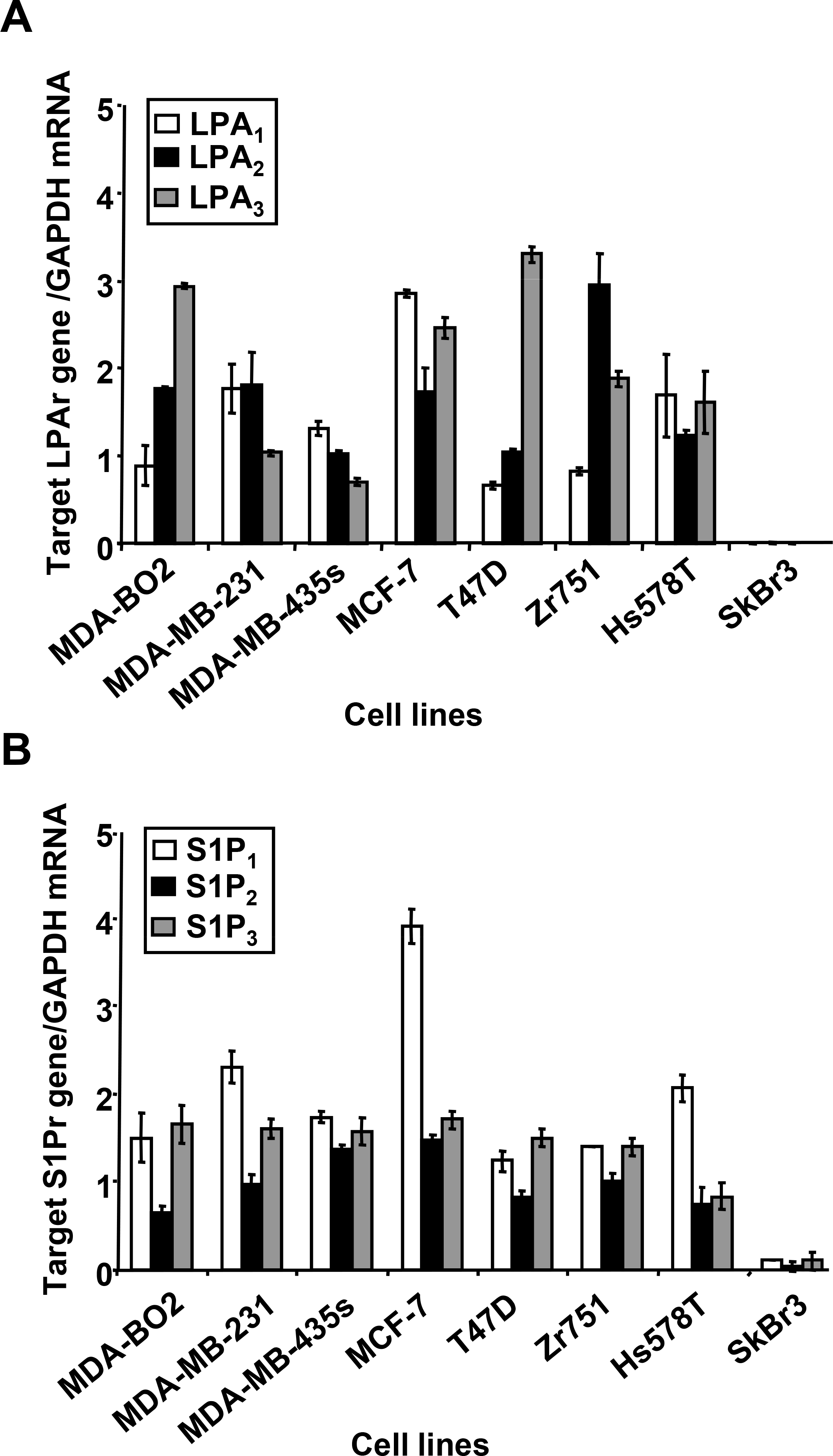
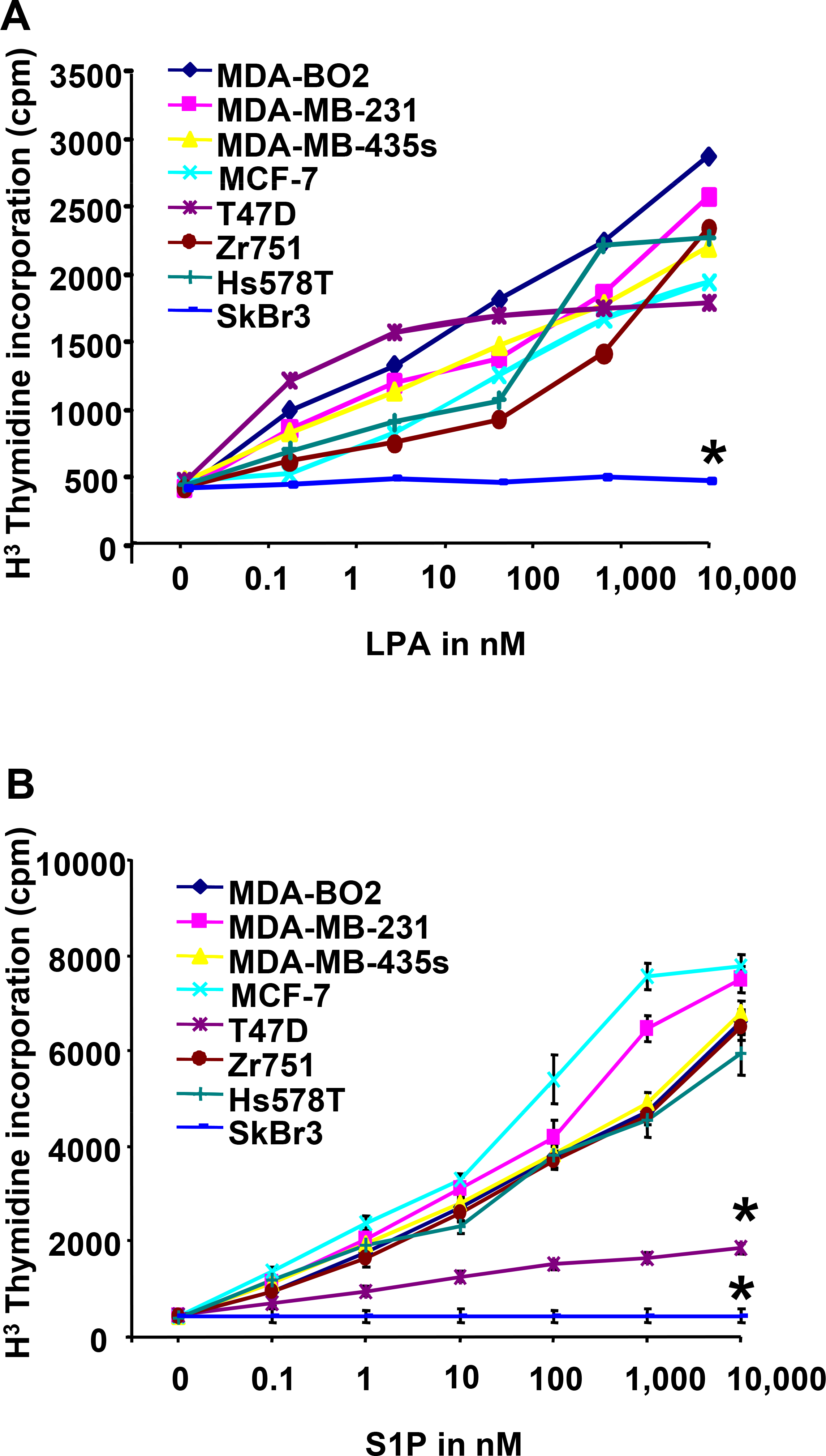
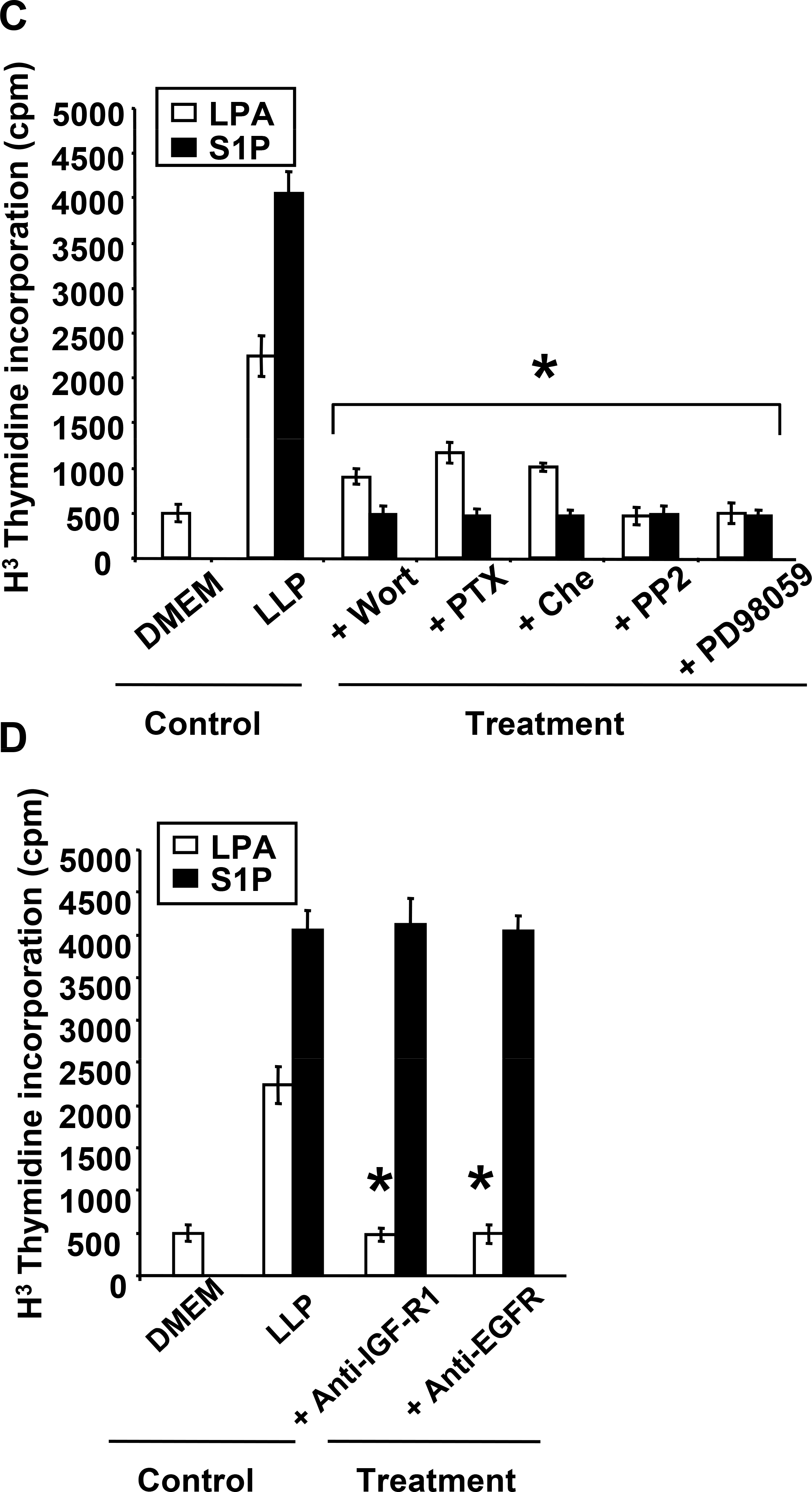
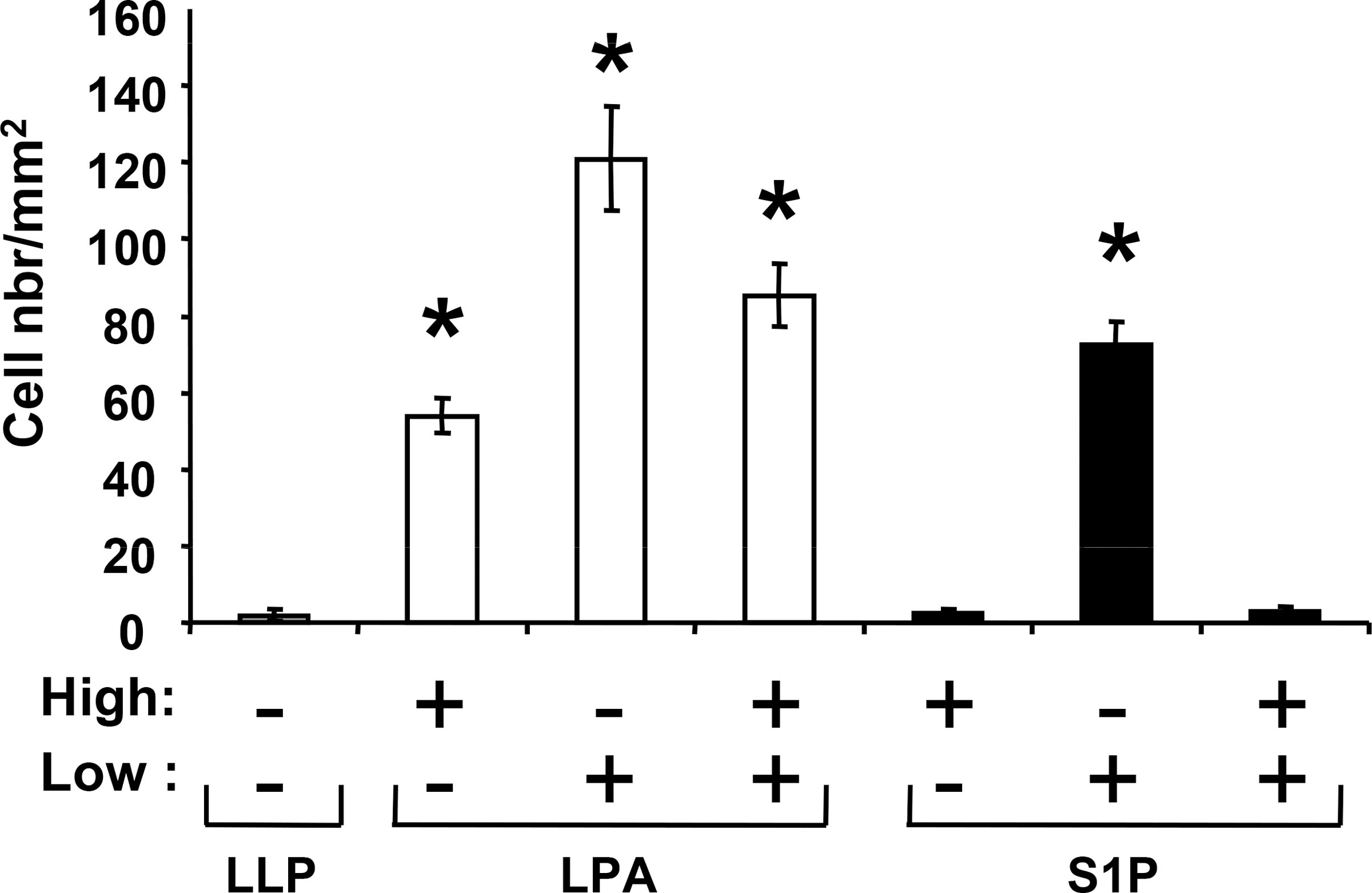
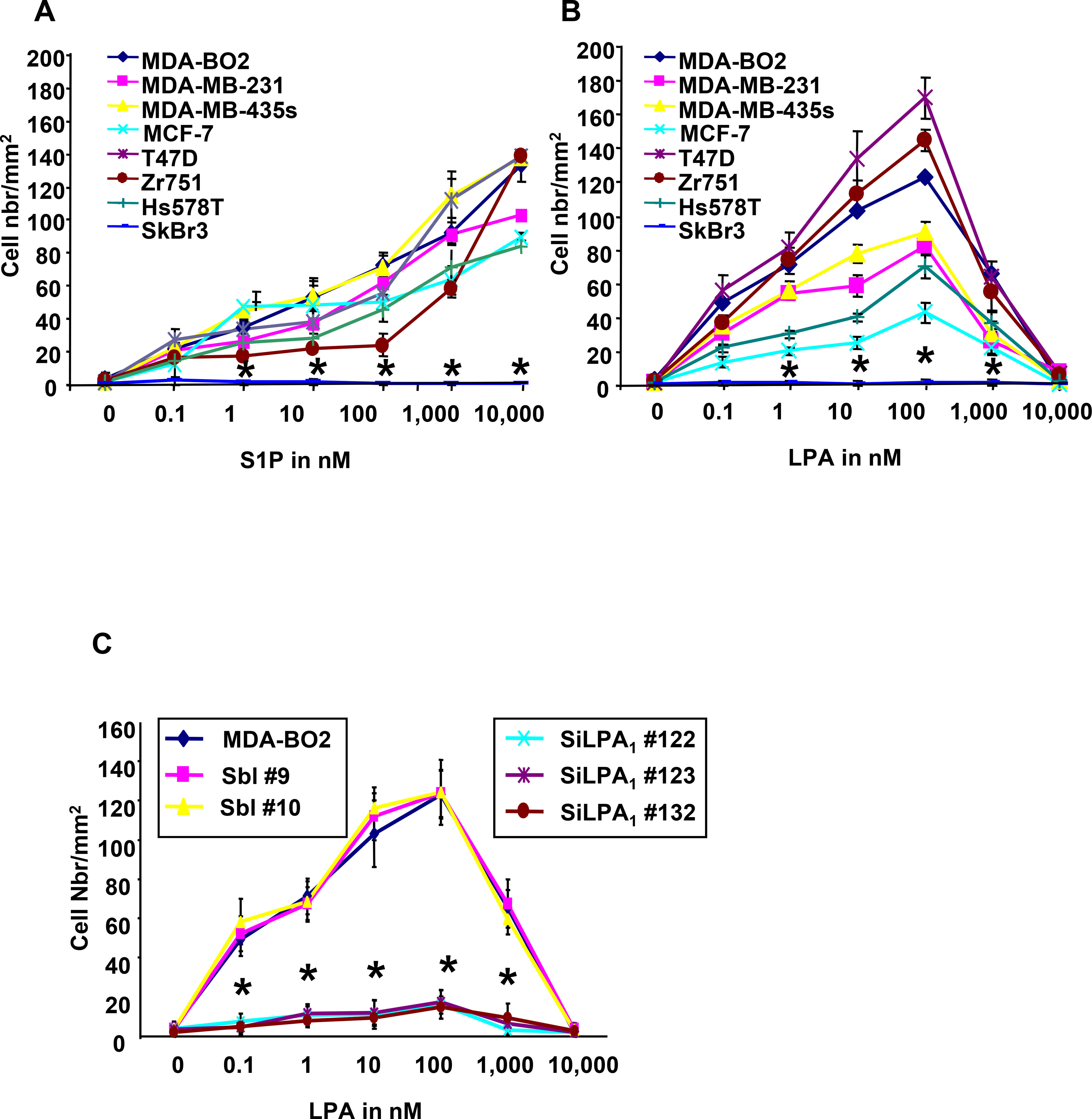
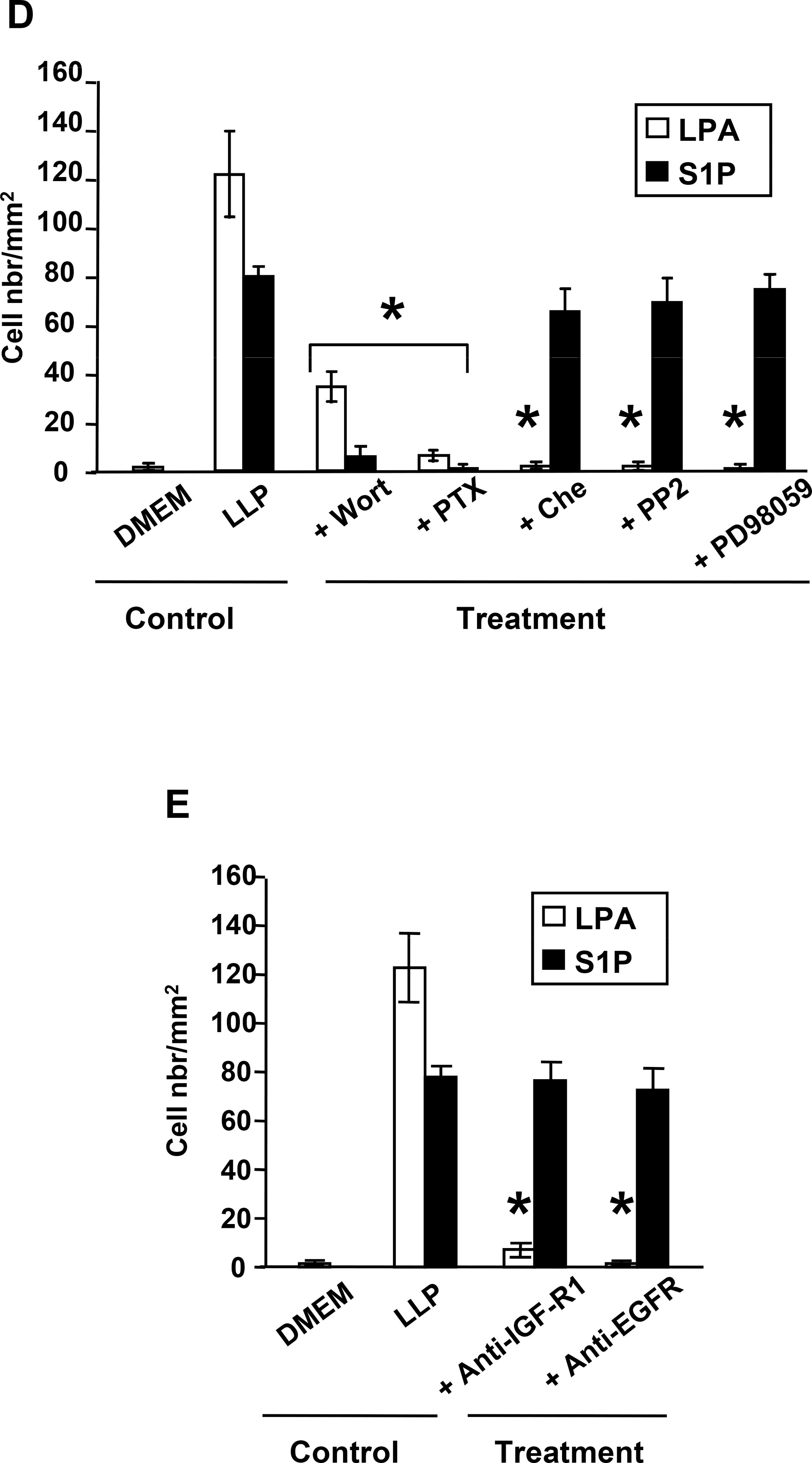
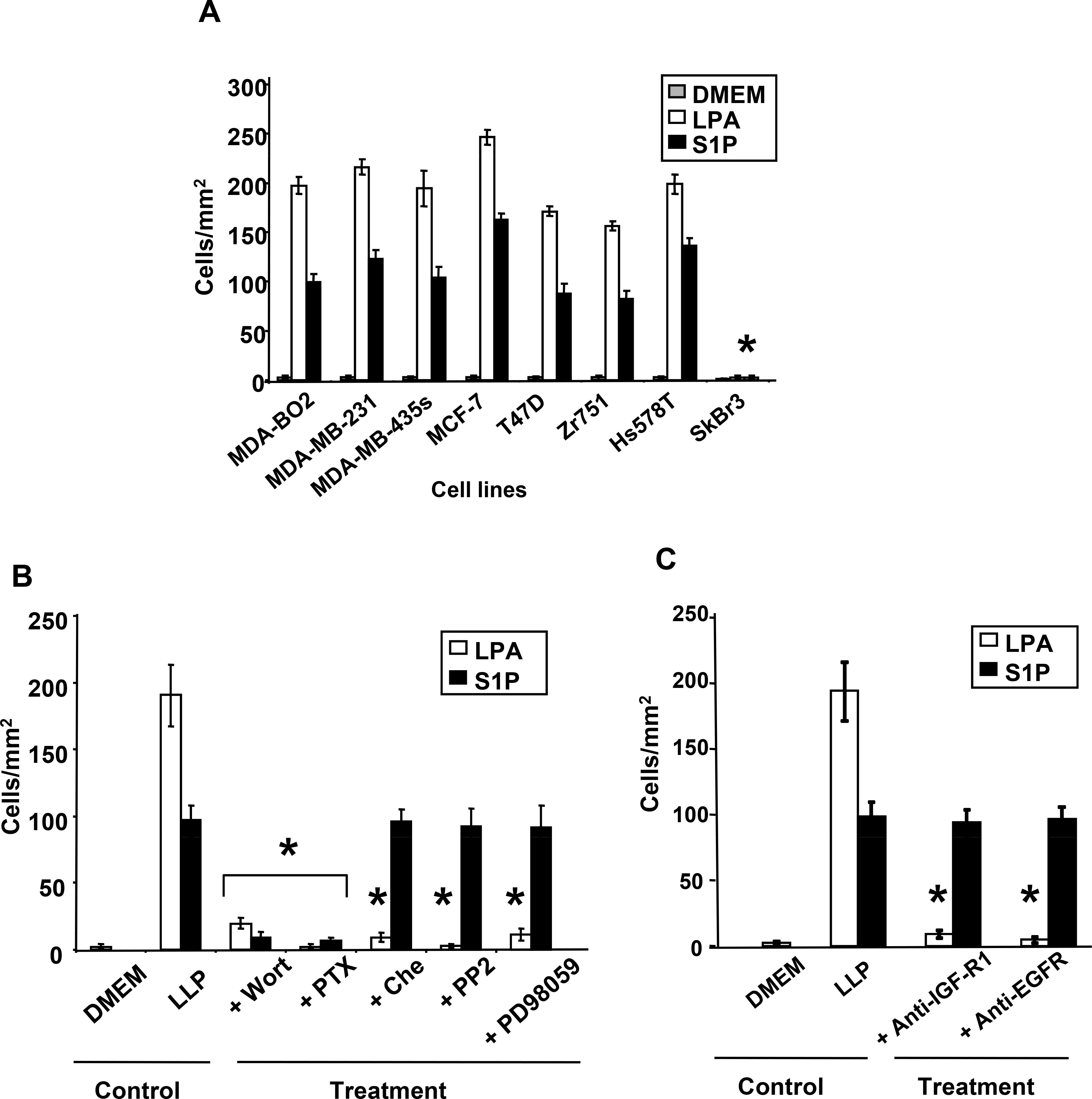
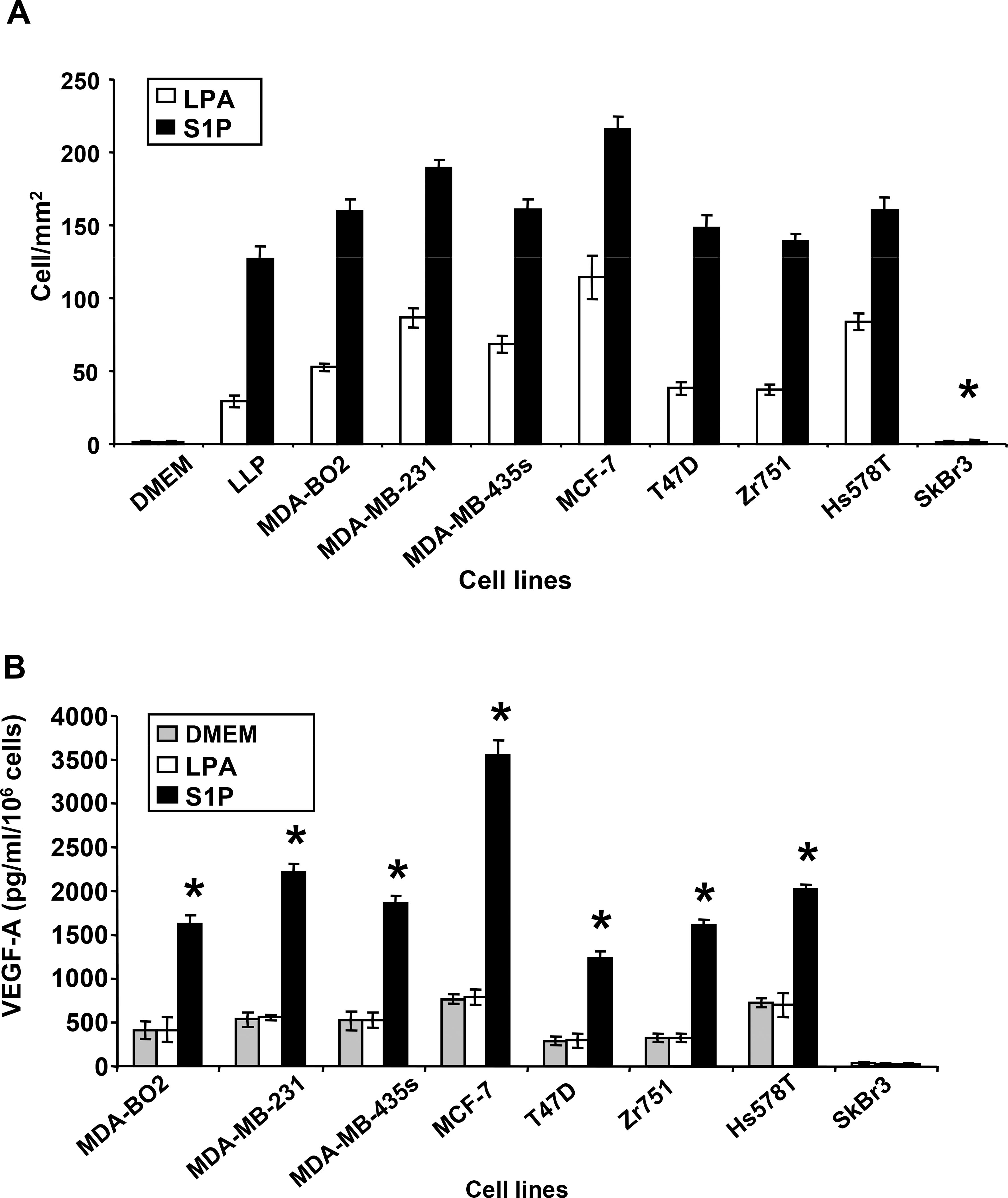
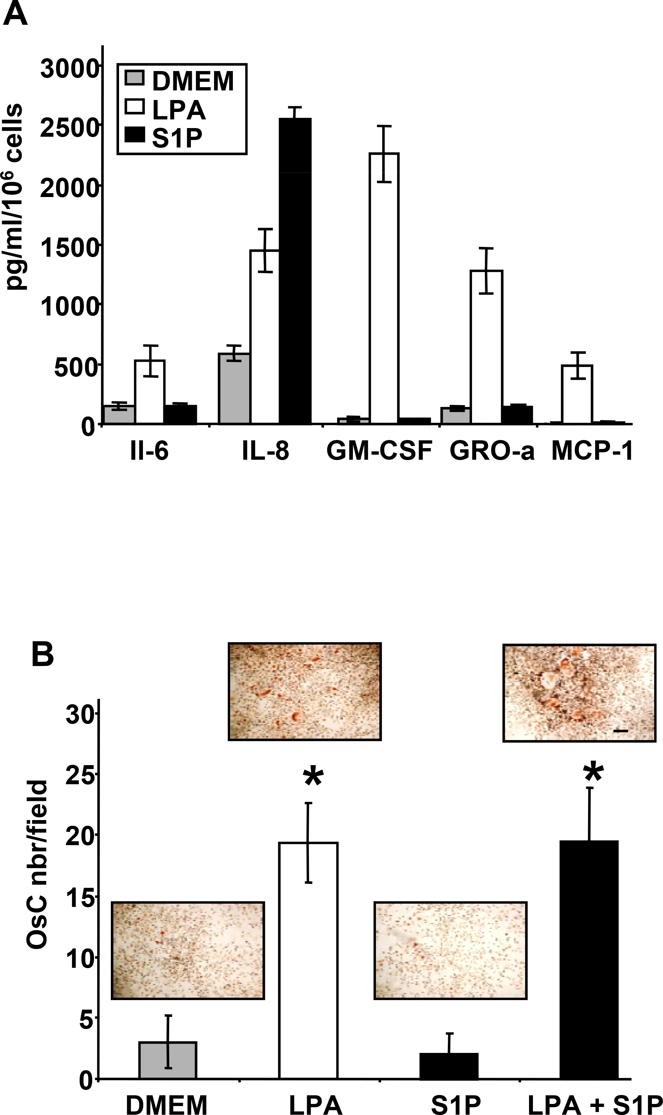
Lysophosphatidic acid (LPA) and sphingosine 1-phosphate (S1P) are structurally related bioactive lipids with growth factor-like activities. LPA and S1P are naturally produced in vivo by blood platelets upon platelet aggregation and at least in vitro by fibroblasts, adipocytes, and multiple types of tumor cells. Breast cancer cells respond to LPA and S1P. However, their specific actions on breast cancer cell biological functions remain unclear. We therefore conducted an in vitro side-by-side study of these two lipids on breast cancer cells. LPA mediates human breast cancer MDA-BO2 cell proliferation, migration, and invasion through activation of a G(alpha i)/ERK1/2-dependent signaling pathway, whereas activation of G(alpha i)/PI3K predominates upon S1P stimulation. In MDA-BO2 cells, LPA but not S1P activities were dependent on active type 1 insulin-like growth factor and epithelial growth factor receptors. LPA and S1P act directly on endothelial cells to induce angiogenesis. We demonstrate that LPA and S1P have indirect angiogenic properties as judged by induced secretion of angiogenic factors by breast cancer cells primed with these lysophospholipids. S1P, but not LPA, controlled the expression of VEGF-A by breast cancer cells, while LPA, but not S1P, controlled the expression of GM-CSF, Gro-alpha, MCP-1, and IL-6. According to the secretion of these paracrine osteoclastic factors, LPA, but not S1P, stimulates breast cancer cell-induced osteoclastogenesis. These findings suggest that, in vivo, LPA and S1P can coordinate their action on tumor and surrounding cells to induce breast cancer progression both at primary and bone metastatic sites.


Figures

Fig. 1–
Fig. 1–. Expression of LPA and S1P receptors in human breast cancer cells lines.
The level of target lysolipid receptor mRNA in breast cancer cell lines was measured using quantitative real-time PCR. Results are expressed as a relative value to GAPDH mRNA. A. Expression levels of LPA receptors (LPA1, LPA2, LPA3). B. Expression levels of S1P receptors (S1P1, S1P2, S1P3). Cell lines are MDA-MB-231, MDA-BO2, MDA-MB-435S, MCF-7, T47D, Zr751, Hs578T and SkBr3. Bars represent means ± SD of at least three experiments.
Fig. 2–
Fig. 2–. LPA and S1P induce breast cancer cell proliferation through distinct signaling pathways.
A. Mitogenic activity of LPA and B. S1P were analyzed on quiescent tumor cell lines treated overnight in the presence of increasing concentrations of either lysolipid (0 to 10−5 M) and pulsed with [3H]-thymidine for the last 8 hours of lysolipid treatment. *, P<0.001 compared to MDA-BO2 cell line. C. Effect of inhibitors PI3K (Wortmannin, 10−7 M), Gαi (PTX, 100ng/mL), PKC (chelerythrine chloride, 10−6 M), Src kinase (PP2, 10−7 M), or ERK1/2 phosphorylation (PD98059, 10−5 M) on the proliferation of MDA-BO2 cells stimulated with either lysolipids (LLP), LPA or S1P (10−7 M). D. Effect of anti-IGFR1 and EGFR antibodies (1μg/ml) on the mitogenic activity of LPA and S1P (10−7 M) on MDA-BO2 cells. Data are expressed as specific H3Thymidine incorporation and represent means ± SD of at least three independent experiments. *, P<0.001 compared to corresponding lysolipid treatment.
Fig. 3–
Fig. 3–. LPA and S1P exhibit differential migratory activities on breast cancer cells.
MDA-BO2 cells were seeded on higher (High) and/or lower (Low) chambers of transwell migration chambers. No lysolipid (LLP) or S1P (10−7M) or LPA (10−7M) were added on indicated chambers. LPA exhibits both chemotactic and chemokinetic activities, whereas S1P acts only as a chemo-attractant factor on MDA-BO2 cells. Cells that have passed through the 8 μm membrane were enumerated after 6 hours of treatment. Data are expressed as number of cells/mm2. Bars represent means ± SD of at least three experiments. *, P<0.001 compared to control condition in absence of lysolipid.
Fig. 4–
Fig. 4–. LPA and S1P induce migration of breast cancer cells through specific signaling pathways.
A and. B. LPA and S1P stimulate dose-dependently the migration of human breast cancer cells that express cognate lysolipid receptors. Increased concentrations (0 to 10−5 M) of S1P (A) or LPA (B) were added in the lower compartments of transwell plate migration chambers. Indicated cell lines were seeded on the higher chamber and challenged to migrate through the filter membrane for 6h. While S1P exhibits a continuous chemotactic activity throughout the range of concentrations, LPA action follows a belt shape curve, with a pic of maximum activity at 10−7M. *, P<0.001 compared to MDA-BO2 cells. C. Silencing of LPA1 expression inhibits LPA-induced migration of MDA-BO2 cells. Three independent SiLPA1 transfectants, two scramble-SiRNA control clones (Sbl) and MDA-BO2 parental cells (3) were incubated with increased concentrations of LPA as described above. *, P<0.001 compared to MDA-BO2 cells. D. Effect of inhibition of signaling pathways on migration of MDA-BO2 cells stimulated with LPA or S1P (10−7M). Inhibitors used were identical as described in Fig. 2B. E. Effect of anti-IGFR1 and EGFR antibodies (1μg/ml) on the migration of MDA-BO2 cells induced by LPA and S1P (10−7 M). Data are expressed in cell number/mm2. Bars represent means ± SD of at least three experiments. *, P<0.001 compared to corresponding lysolipid treatment.
Fig. 5–
Fig. 5–. LPA and S1P stimulate invasion of human breast cancer cells through specific signaling pathways.
Transwell migration chambers were previously coated with Matrigel. A. Cells were incubated in absence (DMEM) or presence of S1P (10−7M) or LPA (10−7M) added in the lower chamber. After 48h hours of lysolipid treatment cells passed thought the matrigel barrier were enumerated. *, P<0.001 compared to MDA-BO2 cells. B. MDA-BO2 cells pretreated with Wortmannin (10−7M), PTX (100ng/mL), chelerythrine chloride (10−6M), PP2 (10−7M), or PD98059 (10−5M) were seeded on the higher chamber and challenged for 48 hours, to invade and migrate through the filter membrane precoated with matrigel, upon stimulation with lysolipids (LPL) added in the lower chamber. C. Effect of anti-IGFR1 and EGFR antibodies (1μg/ml) on invasion of MDA-BO2 cells induced by LPA and S1P (10−7 M). Data are expressed in cell number/mm2. Bars represent means ± SD of at least three experiments. *, P<0.001 compared to corresponding lysolipid treatment.
Fig. 6–
Fig. 6–. Commune induction of tumor cell-mediated angiogenesis by LPA and S1P and specific secretion of VEGF-A by S1P.
A. Conditioned media from human breast cancer cell lines primed with LPA or S1P (10−6M) for 48h, were tested for their capacity to induce HUVEC cell migration. HUVEC cells were seeded in the higher chamber of transwell migration chambers and challenged to migrate through the filter membrane for 6h. Data are expressed as the number of cells per mm2. *, P<0.001 compared to MDA-BO2 cell conditioned medium B. The secretion of VEGF-A was quantified by ELISA from conditioned media collected from indicated breast cancer cell lines stimulated with LPA (10−7 M) or S1P (10−7 M) for 48h. Data are expressed in pg/ml/106 cells. All bars and curves represent means ± SD of at least three experiments. *, P<0.001 compared to DMEM treated cells.
Fig. 7–
Fig. 7–. LPA but not S1P induces pro-osteoclastogenic activity of MDA-BO2 cells.
A. pro-osteoclastogenic factors (IL-6, IL-8, GM-CSF, GRO-a, MCP-1) were quantified by ELISA from conditioned media generated from MDA-BO2 cells stimulated with LPA (10−6M) or S1P (10−6M). B. LPA but not S1P, stimulates tumor cell-induced formation of mature osteoclasts. Bone marrow cells were cultured in the presence of conditioned media collected from MDA-BO2 cells treated with LPA (10−6M) or S1P (10−6M), or with a combination of LPA and S1P (10−6M). Mature osteoclasts, which are TRAP positive multinucleated cells, were enumerated under microscope. Data are expressed as the number of osteoclasts per field. Bars represent means ± SD of at least three experiments. Insets: representative picture of osteoclasts differentiated in each condition. Size ladder 100 μm. *, P<0.001 compared to DMEM treated cells.

Similar articles

Cited by

References

    1. Boucharaba A; Serre CM; Gres S; Saulnier-Blache JS; Bordet JC; Guglielmi J; Clezardin P; and Peyruchaud OJ Clin. Invest. 114: 1714–1725. 2004. - PMC - PubMed
    1. Visentin B; Vekich JA; Sibbald BJ; Cavalli AL; Moreno KM; Matteo RG; Garland WA; Lu Y; Yu S; Hall HS; Kundra V; Mills GB; and Sabbadini RA Cancer Cell 9: 225–238. 2006. - PubMed
    1. Boucharaba A; Serre CM; Guglielmi J; Bordet JC; Clezardin P; and Peyruchaud O Proc Natl Acad Sci U S A 103: 9643–9648. 2006. - PMC - PubMed
    1. Ishii I; Fukushima N; Ye X; and Chun J Annu Rev Biochem 73: 321–354. 2004. - PubMed
    1. Birgbauer E; and Chun J Cell. Mol. Life Sci. 63: 2695–2701. 2006. - PMC - PubMed

Publication types

MeSH terms