

Development of a catheter for combined intravascular ultrasound and photoacoustic imaging
- PMID: 20113121
- PMCID: PMC2814830
- DOI: 10.1063/1.3274197
Development of a catheter for combined intravascular ultrasound and photoacoustic imaging
Abstract
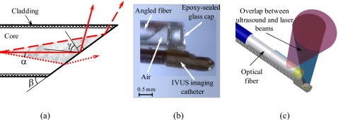
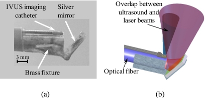
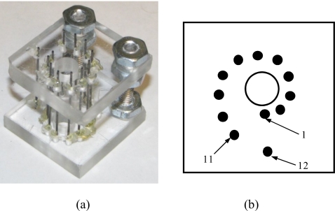
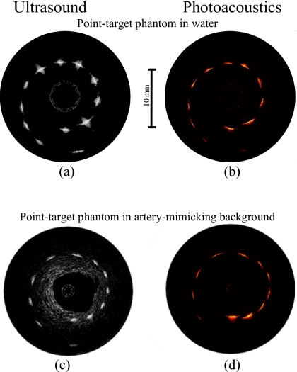
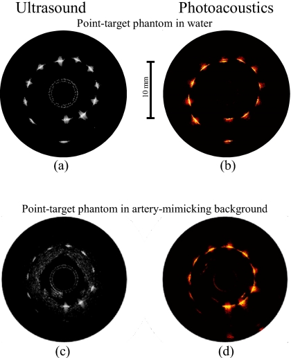
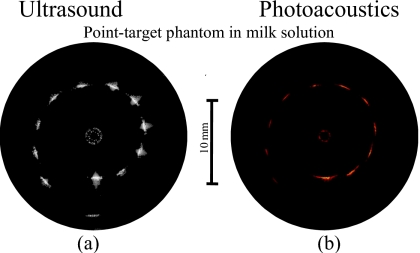
Atherosclerosis is characterized by formation and development of the plaques in the inner layer of the vessel wall. To detect and characterize atherosclerotic plaques, we previously introduced the combined intravascular ultrasound (IVUS) and intravascular photoacoustic (IVPA) imaging capable of assessing plaque morphology and composition. The utility of IVUS/IVPA imaging has been demonstrated by imaging tissue-mimicking phantoms and ex vivo arterial samples using laboratory prototype of the imaging system. However, the clinical realization of a IVUS/IVPA imaging requires an integrated intravascular imaging catheter. In this paper, two designs of IVUS/IVPA imaging catheters--side fire fiber-based and mirror-based catheters--are reported. A commercially available IVUS imaging catheter was utilized for both pulse-echo ultrasound imaging and detection of photoacoustic transients. Laser pulses were delivered by custom-designed fiber-based optical systems. The optical fiber and IVUS imaging catheter were combined into a single device. Both designs were tested and compared using point targets and tissue-mimicking phantoms. The results indicate applicability of the proposed catheters for clinical use.
Figures
References
-
- Falk E., Shah P. K., and Fuster V., Circulation 92, 657 (1995). - PubMed
-
- Naghavi M., Libby P., Falk E., Casscells S. W., Litovsky S., Rumberger J., Badimon J. J., Stefanadis C., Moreno P., Pasterkamp G., Fayad Z., Stone P. H., Waxman S., Raggi P., Madjid M., Zarrabi A., Burke A., Yuan C., Fitzgerald P. J., Siscovick D. S., de Korte C. L., Aikawa M., Airaksinen K. E. J., Assmann G., Becker C. R., Chesebro J. H., Farb A., Galis Z. S., Jackson C., Jang I. -K., Koenig W., Lodder R. A., March K., Demirovic J., Navab M., Priori S. G., Rekhter M. D., Bahr R., Grundy S. M., Mehran R., Colombo A., Boerwinkle E., Ballantyne C., W.Insull, Jr., Schwartz R. S., Vogel R., Serruys P. W., Hansson G. K., Faxon D. P., Kaul S., Drexler H., Greenland P., Muller J. E., Virmani R., Ridker P. M., Zipes D. P., Shah P. K., and Willerson J. T., Circulation 108, 1664 (2003). 10.1161/01.CIR.0000087480.94275.97 - DOI - PubMed
-
- Stary H. C., Chandler A. B., and Dinsmore R. E., Arterioscler. Thromb. Vasc. Biol. 15, 1512 (1995). - PubMed
Publication types
MeSH terms
Grants and funding
LinkOut - more resources
Full Text Sources