Comparative Study
. 2010 Mar;29(3):103-20.
doi: 10.1089/dna.2009.0963.

A confocal and electron microscopic comparison of interferon beta-induced changes in vesicular stomatitis virus infection of neuroblastoma and nonneuronal cells

Affiliations
Comparative Study

A confocal and electron microscopic comparison of interferon beta-induced changes in vesicular stomatitis virus infection of neuroblastoma and nonneuronal cells

Paul M D'Agostino et al. DNA Cell Biol. 2010 Mar.

Abstract

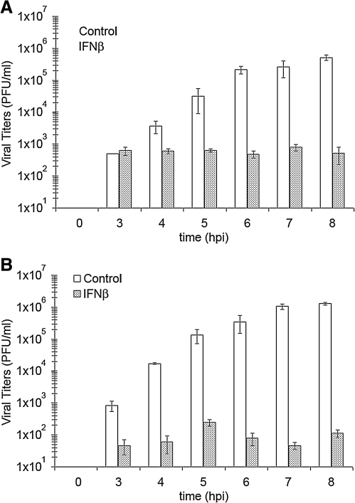
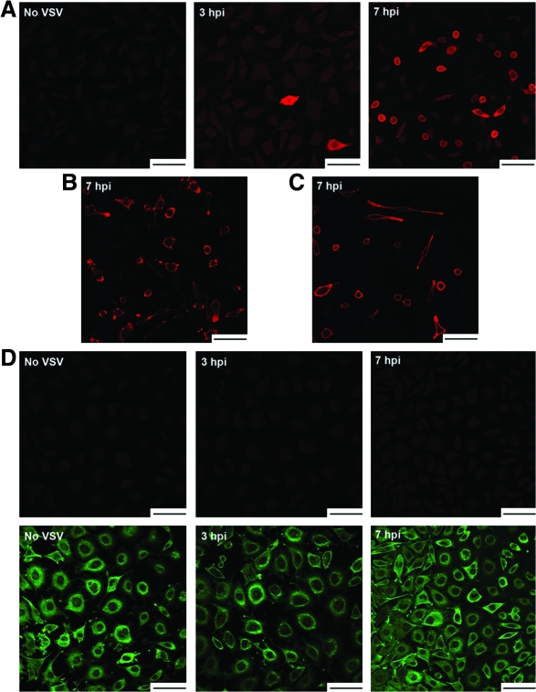
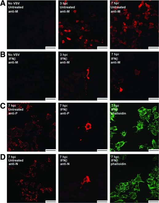
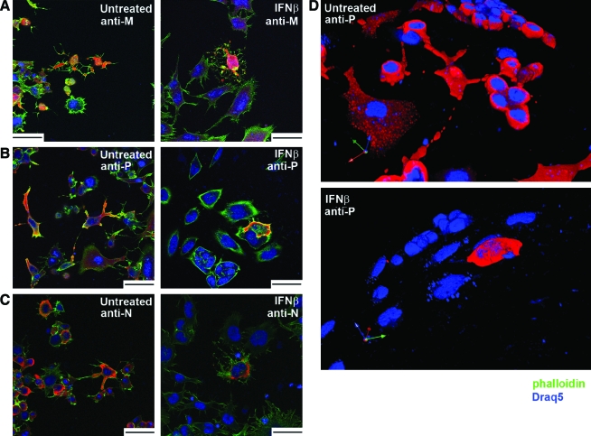
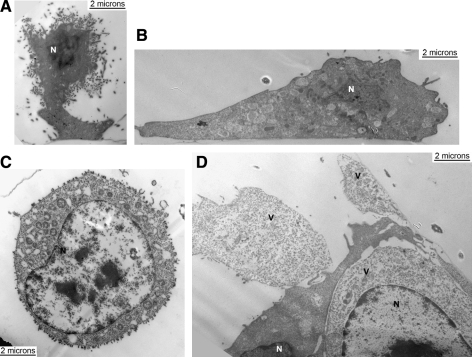
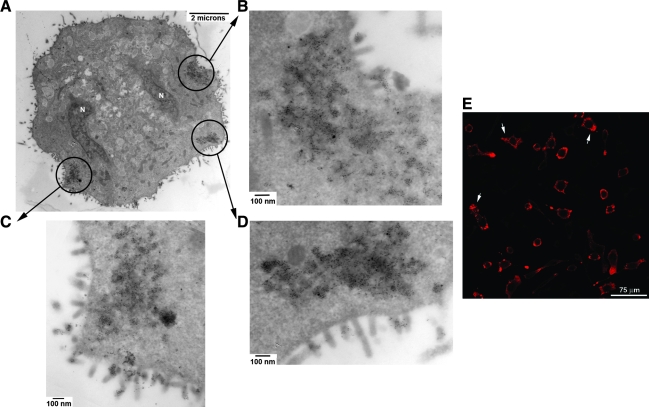
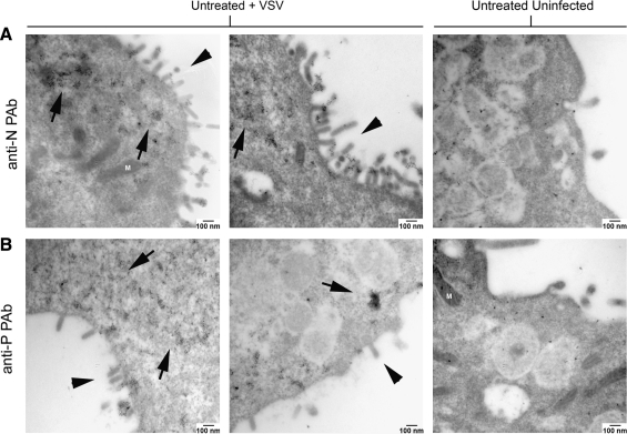
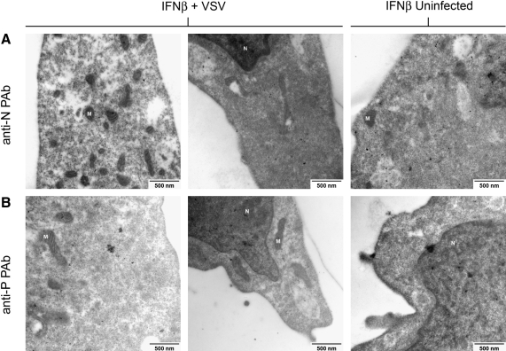
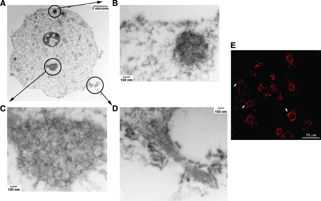
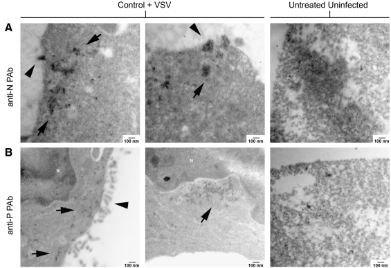
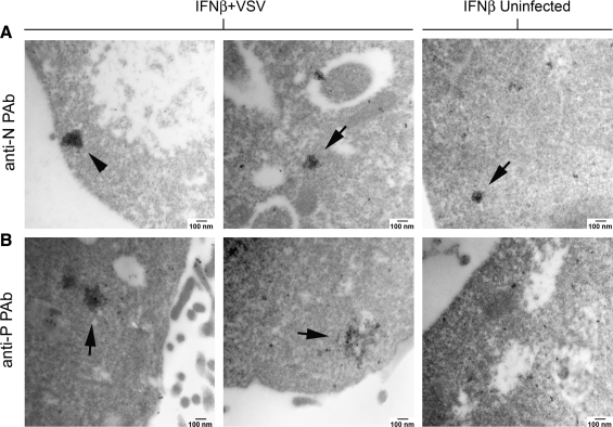
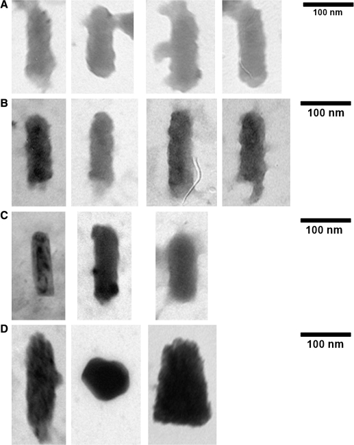
Vesicular stomatitis virus (VSV) replication is highly sensitive to interferon (IFN)-induced antiviral responses. Pretreatment of sensitive cultured cells with IFNbeta results in a 10(4)-fold reduction in the release of infectious VSV particles. However, differences exist between the mechanisms of reduced infectious particle titers in cell lines of neuroblastoma and nonneuronal lineage. In L929-fibroblast-derived cells, using immunofluorescence confocal microscopy, infection under control conditions reveals the accumulation of VSV matrix, phosphoprotein (P), and nucleocapsid (N) proteins over time, with induced cellular morphological changes indicative of cytopathic effects (CPEs). Upon observing L929 cells that had been pretreated with IFNbeta, neither detectable VSV proteins nor CPEs were seen, consistent with type I IFN antiviral protection. When using the same techniques to observe VSV infections of NB41A3 cells, a neuroblastoma cell line, aside from similar viral progression in the untreated control cells, IFNbeta-treated cells illustrated a severely attenuated VSV infection. Attenuated VSV progression was observed through detection of VSV matrix, P, and N proteins in isolated cells during the first 8 h of infection. However, by 18-24 h postinfection all neuroblastomas had succumbed to the viral infection. Finally, upon closer inspection of IFNbeta-treated NB41A3 cells, no detectable changes in VSV protein localization were identified compared with untreated, virally infected neuroblastomas. Next, to extend our study to test our hypothesis that virion assembly is compromised within type I IFN-treated neuroblastoma cells, we employed electron microscopy to examine our experimental conditions at the ultrastructural level. Using VSV-specific antibodies in conjunction with immuno-gold reagents, we observed several similarities between the two cell lines, such as identification of viroplasmic regions containing VSV N and P proteins and signs of stress-induced CPEs of VSV-infected cells, which had either been mock-treated or pretreated with interferon-beta (IFNbeta). One difference we observed between nonneuronal and neuroblastoma cells was more numerous actively budding VSV virions across untreated L929 plasma membranes compared with untreated NB41A3 cells. Additionally, IFNbeta-treated, VSV-infected L929 cells exhibited neither cytoplasmic viroplasm nor viral protein expression. In contrast, IFNbeta-treated, VSV-infected NB41A3 cells showed evidence of VSV infection at a very low frequency as well as small-scale viroplasmic regions that colocalized with viral N and P proteins. Finally, we observed that VSV viral particles harvested from untreated VSV-infected L929 and NB41A3 cells were statistically similar in size and shape. A portion of VSV virions from IFNbeta-treated, virally infected NB41A3 cells were similar in size and shape to virus from both untreated cell types. However, among the sampling of virions, pleomorphic viral particles that were identified from IFNbeta-treated, VSV-infected NB41A3 cells were different enough to suggest a misassembly mechanism as part of the IFNbeta antiviral state in neuroblastoma cells.


Figures

FIG. 1.
FIG. 1.
Infectious VSV particles released from neuronal and nonneuronal cells during an 8 h time course. Supernatants were harvested from VSV-infected (multiplicity of infection = 3) (A) L929 and (B) NB41A3 cells either pretreated with 400 U/mL IFNβ or vehicle alone at various time points during an 8 h in vitro infection and assessed by plaque assay. In both cell lines viral titers steadily increased in untreated samples, but remained at background levels under IFNβ-treated conditions. Samples were assayed in triplicate during three separate experiments, and error bars represent 2 × standard deviation. VSV, vesicular stomatitis virus; IFNβ, interferon β.
FIG. 2.
FIG. 2.
VSV progression through nonneuronal L929 cells during an 8 h time course and the IFNβ effect on that progression. VSV protein expression was seen steadily increasing in greater numbers throughout the course of infection in untreated L929 cells by observing cells stained with (A) anti-VSV M antibody (red), (B) anti-VSV P antibody (red), or (C) anti-VSV N antibody (red). (D) No viral protein expression was observed in IFNβ-pretreated L929 cells stained with phalloidin (green) and anti-VSV M antibody (red). Staining of the entire time course in untreated and IFNβ-pretreated L929 cells was not included for clarity and space considerations. Draq5 nuclear staining was also omitted for clarity. This figure is representative of three independently conducted experiments. Scale bars = 75 μm. M, matrix; N, nucleocapsid; P, phosphoprotein.
FIG. 3.
FIG. 3.
VSV viral protein expression patterns in IFNβ-induced NB41A3 neuroblastoma varies greatly from that in similarly treated L929 cells. VSV-infected, (A) untreated and (B) IFNβ-pretreated NB41A3 cells were stained with anti-VSV M antibody (red). Analogous to untreated L929 cells, untreated NB41A3 cells showed increased VSV M expression during the course of an 8 h infection (only partially shown for brevity). However, unlike what was observed in IFNβ-pretreated L929 cells, in IFNβ-induced NB41A3 cells VSV M protein expression was detected in a very small fraction of virally infected cells (roughly 1:10,000 cells) (also only partially shown for brevity). The positively stained fraction observed a slow increase during the infection time course with adjacent cells expressing the VSV M protein. When allowed to proceed for greater than 24 hpi, all cells succumbed to the VSV infection. (C) In addition, VSV-infected, untreated (left panel) and IFNβ-pretreated NB41A3 cells (middle and right panels) were stained with anti-VSV P antibody (red; left and middle panels) and phalloidin (green; right panel). (D) Similar to (C) except that cells were stained with anti-VSV N antibody (red; left and middle panels). Positively stained fractions observed a slow increase during infections with adjacent cells expressing the VSV P or N proteins. Draq5 nuclear staining was omitted for clarity; this figure is representative of three independently conducted experiments. Scale bars = 75 μm.
FIG. 4.
FIG. 4.
The VSV protein localization in IFNβ-induced NB41A3 neuroblastomas is no different from its distribution in untreated cells. (A) VSV-infected, untreated and IFNβ-pretreated NB41A3 cells were stained with phalloidin (green), Draq5 (blue), and anti-VSV M antibody (red). Using the Leica AF software package, stacked images were exported as .avi movie files to observe cell sections, through the z-plane, to identify the VSV M protein cellular localization pattern in untreated (left panel) and IFNβ-pretreated NB41A3 cells (right panel). (B) Similar analysis of VSV-infected, untreated and IFNβ-pretreated NB41A3 cells were stained with phalloidin (green), Draq5 (blue), and anti-VSV P antibody (red). (C) Representative image from VSV-infected, untreated and IFNβ-pretreated NB41A3 cells were stained with phalloidin (green), Draq5 (blue), and anti-VSV N antibody (red). (D) To verify what was observed via sectional analysis and to pick up any three-dimensional patterns not represented in the two-dimensional sections, stacked z-plane images were projected into three dimensions using the Volocity software package to further aid in observing the VSV protein localization patterns. An example of such a projection is shown for anti-VSV P staining in untreated (upper panel) and IFNβ-pretreated cells (lower panel). Both revealed not changes in viral protein localization due to an IFNβ-induced antiviral state in the neuroblastoma cell line. This figure is representative of three independently conducted experiments. Scale bars = 50 μm.
FIG. 5.
FIG. 5.
General observations on VSV-infected L929 and NB41A3 cells with and without IFNβ treatment at 18–24 hpi. (A) An untreated L929 cell infected with VSV experiencing cytopathic effects (CPEs; sagittal view in relation to the extracellular matrix). (B) An IFNβ-treated L929 cell infected with VSV (sagittal view). (C) An untreated NB41A3 cell infected with VSV with a swollen nucleus (transverse view). (D) Four IFNβ-treated NB41A3 cells infected with VSV, in which three cells, labeled “V,” are experiencing CPEs from the infection (transverse view). This figure is representative of three independently conducted experiments; N denotes nuclei.
FIG. 6.
FIG. 6.
Untreated, VSV-infected L929 cells have electron-dense regions containing viral proteins at 6 hpi. (A) Electron micrograph at 10,000 × magnification. Circled areas denote viroplasmic regions that are enlarged to 75,000 × in (B–D), where colocalization of VSV P protein is observed by the collection of 6 nm gold particles. (E) Confocal image from VSV-infected L929 cells stained with anti-N antiserum (red) showing aggregate staining (white arrows) within the cytoplasm that may correlate with the same viroplasmic regions detected by electron microscope. This figure is representative of three independently conducted experiments; N represents the nucleus.
FIG. 7.
FIG. 7.
Viroplasm-like regions within untreated, VSV-infected L929 cells contain both VSV P and N proteins. (A) Untreated, VSV-infected L929 cells at 6 hpi or untreated, uninfected L929 cells were probed with rabbit anti-VSV N antiserum. (B) Likewise untreated, VSV-infected L929 cells at 6 hpi or untreated, uninfected L929 cells were probed with rabbit anti-VSV P antiserum. Samples were subsequently treated with goat anti-rabbit immunoglobulin G-conjugated with 6 nm gold particles. Electron micrographs at 75,000 × magnification show viroplasmic regions colocalized with VSV N and P proteins, as observed by the collection of 6 nm gold particles near regions of active virion budding. The level of anti-VSV P staining (on average 469 ± 144 gold particles per 75,000 × viewing field) was well above background (47 ± 9 gold particles per 75,000 × viewing field). Similarly, the levels of VSV N staining (237 ± 53 gold particles per 75,000 × viewing field) were also well over background (51 ± 6 gold particles per 75,000 × viewing field). This figure is representative of three independently conducted experiments; arrows denote electron-dense viroplasmic areas, arrow heads represent budding VSV, and M highlights mitochondria.
FIG. 8.
FIG. 8.
Viroplasmic regions are not found in IFNβ-treated, VSV-infected L929 cells at 6 hpi. IFNβ-treated, VSV-infected L929 cells at 6 hpi and IFNβ-treated, uninfected L929 cells were probed in a similar fashion as the cells in Figure 7. After examining 20–30 electron micrographs per treatment group, at 75,000 × magnification, neither electron-dense regions nor the expression of (A) VSV N (83 ± 35 gold particles for anti-N staining in IFNβ-treated, VSV-infected cells versus 76 ± 13 for anti-N staining in IFNβ-treated, uninfected cells in a 75,000 × viewing field) and VSV P proteins were identified (49 ± 9 gold particles for anti-P staining in IFNβ-treated, VSV-infected cells versus 45 ± 2 for anti-P staining in IFNβ-treated, uninfected cells in a 75,000 × viewing field). This figure is representative of three independently conducted experiments; N denotes nuclei and M highlights mitochondria.
FIG. 9.
FIG. 9.
Untreated, VSV-infected NB41A3 cells show viroplasm-like regions containing viral proteins at 6 hpi. (A) Untreated, VSV-infected NB41A3 cells were probed with rabbit anti-VSV P antiserum and secondary goat anti-rabbit immunoglobulin G–conjugated with 6 nm gold particles. Circled areas denote electron-dense regions that are enlarged to 75,000 × in (B) and (C), where colocalization of VSV P protein is observed by the collection of 6 nm gold particles. (D) Depicts an area of active viral budding. (E) Confocal image from VSV-infected NB41A3 cells stained with anti-N antiserum (red) showing aggregate staining (white arrows) within the cytoplasm that may correlate with the same viroplasmic regions detected by electron microscope. This figure is representative of three independently conducted experiments; N represents the nucleus.
FIG. 10.
FIG. 10.
Viroplasmic regions within untreated, VSV-infected NB41A3 cells also contain both VSV P and N proteins. Untreated, uninfected or untreated, VSV-infected NB41A3 cells were stained as described in Figure 7. Electron micrographs at 75,000 × magnification show viroplasm-like regions colocalized with VSV N and P proteins, as observed by the collection of 6 nm gold particles. Unlike L929 cells, these viroplasmic regions were not near regions of active virion budding and were not as heavily electron dense. (A) The levels of anti-VSV N (on average 174 ± 41 gold particles per 75,000 × viewing field) and (B) P staining (264 ± 98 gold particles per 75,000 × viewing field) were well above background (respectively, 48 ± 17 gold particles and 47 ± 7 gold particles per 75,000 × viewing field). This figure is representative of three independently conducted experiments; arrows denote viroplasmic areas, arrow heads represent budding VSV, and N highlights nuclei.
FIG. 11.
FIG. 11.
Unlike L929 cells, small viroplasmic regions are found in IFNβ-treated, VSV-infected NB41A3 cells and contain both VSV P and N proteins at 6 hpi. IFNβ-treated, VSV-infected NB41A3 cells at 6 hpi or IFNβ-treated uninfected were again stained as in Figure 7. Electron micrographs at 75,000 × magnification show substantially smaller electron dense regions colocalized with either (A), VSV N or (B) VSV P proteins. The viroplasmic regions were sometimes so small (82 ± 28 gold particles for anti-P staining and 103 ± 9 for anti-N staining in a 75,000 × viewing field) and they were difficult to discern from background staining (respectively, 43 ± 11 gold particles for anti-P staining and 99 ± 13 for anti-N staining in a 75,000 × viewing field) in IFNβ-treated, uninfected cells (arrow head, upper right panel). Despite the appearance of viroplasm-like regions, no actively budding VSV was observed. This figure is representative of three independently conducted experiments; arrows denote viroplasmic areas.
FIG. 12.
FIG. 12.
VSV viral particles that budded from IFNβ-treated NB41A3 cells contained characteristic bullet shape as well as pleomorphic virion-like particles. Supernatants from VSV infections of untreated NB41A3 or L929 cells were harvested at 10 hpi and supernatants from IFNβ-treated cells were harvested at 18–24 hpi. Viral particles were purified and then fixed then stained with 1% osmium tetroxide in 100 mM cacodylate buffer pH 7.2 and 4% uranyl acetate. Electron micrographs at 100,000 × of VSV viral particles from (A) untreated, VSV-infected L929 cells and (B) untreated, VSV-infected NB41A3 cells show no statistical difference in virion length or width; the same was true of many virions from (C) IFNβ-treated, VSV-infected NB41A3 cells that could be recognized as VSV viral particles. (D) However, another group of particles only observed in crude VSV preparations from IFNβ-treated, VSV-infected NB41A3 cells possessed uncharacteristic, pleomorphic appearance that were either too big or a completely different morphology from what is expected from a Rhabdovirus. Therefore, without a reproducible means of identification these shapes cannot be deemed VSV virions with any certainty, but were consistent with misassembly or an increase in defective particles. This figure is representative of three independently conducted experiments with a total sample size of n = 30–40 for virions from untreated cells; due to restrictions on viral yield from IFNβ-treated cells, sample sizes were n = 5.

Similar articles

Cited by

References

    1. Aloisi F. Immune function of microglia. Glia. 2001;36:165–179. - PubMed
    1. Argyris E.G. Acheampong E. Wang F. Huang J. Chen K. Mukhtar M. Zhang H. The interferon-induced expression of APOBEC3G in human blood-brain barrier exerts a potent intrinsic immunity to block HIV-1 entry to central nervous system. Virology. 2007;367:440–451. - PMC - PubMed
    1. Barr J.N. Whelan S.P. Wertz G.W. Transcriptional control of the RNA-dependent RNA polymerase of vesicular stomatitis virus. Biochim Biophys Acta. 2002;1577:337–353. - PubMed
    1. Belkowski L.S. Sen G.C. Inhibition of vesicular stomatitis viral mRNA synthesis by interferons. J Virol. 1987;61:653–660. - PMC - PubMed
    1. Blondel D. Harmison G.G. Schubert M. Role of matrix protein in cytopathogenesis of vesicular stomatitis virus. J Virol. 1990;64:1716–1725. - PMC - PubMed

Publication types

MeSH terms