. 2010 Feb;31(3):463-76.
doi: 10.1111/j.1460-9568.2009.07058.x. Epub 2010 Jan 26.

Nicotine facilitates long-term potentiation induction in oriens-lacunosum moleculare cells via Ca2+ entry through non-alpha7 nicotinic acetylcholine receptors

Affiliations

Nicotine facilitates long-term potentiation induction in oriens-lacunosum moleculare cells via Ca2+ entry through non-alpha7 nicotinic acetylcholine receptors

Yousheng Jia et al. Eur J Neurosci. 2010 Feb.

Abstract

Hippocampal inhibitory interneurons have a central role in the control of network activity, and excitatory synapses that they receive express Hebbian and anti-Hebbian long-term potentiation (LTP). Because many interneurons in the hippocampus express nicotinic acetylcholine receptors (nAChRs), we explored whether exposure to nicotine promotes LTP induction in these interneurons. We focussed on a subset of interneurons in the stratum oriens/alveus that were continuously activated in the presence of nicotine due to the expression of non-desensitizing non-alpha7 nAChRs. We found that, in addition to alpha2 subunit mRNAs, these interneurons were consistently positive for somatostatin and neuropeptide Y mRNAs, and showed morphological characteristics of oriens-lacunosum moleculare cells. Activation of non-alpha7 nAChRs increased intracellular Ca(2+) levels at least in part via Ca(2+) entry through their channels. Presynaptic tetanic stimulation induced N-methyl-D-aspartate receptor-independent LTP in voltage-clamped interneurons at -70 mV when in the presence, but not absence, of nicotine. Intracellular application of a Ca(2+) chelator blocked LTP induction, suggesting the requirement of Ca(2+) signal for LTP induction. The induction of LTP was still observed in the presence of ryanodine, which inhibits Ca(2+) -induced Ca(2+) release from ryanodine-sensitive intracellular stores, and the L-type Ca(2+) channel blocker nifedipine. These results suggest that Ca(2+) entry through non-alpha7 nAChR channels is critical for LTP induction. Thus, nicotine affects hippocampal network activity by promoting LTP induction in oriens-lacunosum moleculare cells via continuous activation of non-alpha7 nAChRs.


Figures
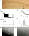
Figure 1
Figure 1. Horizontally oriented interneurons at the stratum oriens/alveus border are nicotine-sensitive
(A) Localization of α2 mRNA-containing interneurons in the hippocampal CA1 region by non-radioactive in situ hybridization. (B1) Bath application of nicotine (10 μM) in the presence of DNQX (20 μM) and AP5 (40 μM) increased the frequency of action potentials in current-clamped oriens/alveus interneurons, which was blocked by the non-α7 nAChR antagonist DHβE (2 μM). (B2) Summary plot of the effect of nicotine (10 μM) on the frequency of action potentials. The nicotine-induced change in the frequency of action potentials was normalized as a percentage change from the highest frequency of action potentials and was plotted against time. (B3) Summary plot of the effect of nicotine (10 μM) on the frequency of action potentials (mean ± SEM) in the absence and presence of DHβE (2 μM). Numbers in parentheses in this and the following figures indicate the numbers of experiments. (C1, C2) IR-DIC images of nicotine-sensitive oriens/alveus interneurons. These interneurons were covered by perineuronal glial cells (C1, the plane of focus was at the neuron under glial cells) or associated with perineuronal glial cells (C2). ***P < 0.001.
Figure 2
Figure 2. A subtype of nicotine-sensitive oriens/alveus interneurons and its molecular markers
(A1, A2) Nicotine-sensitive interneurons contain somatostatin and neuropeptide Y mRNAs. (A1, top) RT-multiplex PCR amplification of different marker sequences. Hippocampal RNA (500 pg) was subjected to a RT-multiplex PCR protocol to detect the expression of calbindin D28k (CB), parvalbumin (PV), calretinin (CR), neuropeptide Y (NPY), vasoactive intestinal polypeptide (VIP), somatostatin (SS), and cholecystokinin (CCK). (A1, bottom) RT-multiplex PCR applied on a single nicotine-sensitive interneuron after electrophysiological recording to detect molecular markers. An example of single-cell RT-multiplex PCR, showing the presence of CR, NPY, VIP, and SS in a recorded cell. RT-PCR products were separated by agarose gel electrophoresis and visualized with ethidium bromide. The amplified fragments had the sizes (in bp) predicted by the mRNA sequences: 432 (CB), 388 (PV), 309 (CR), 359 (NPY), 287 (VIP), 209 (SS), 216 (CCK). A 100 bp DNA ladder was used as a molecular weight marker. (A2) Summary data of single-cell RT-multiplex PCR. (B) One biocytin-labeled nicotine-sensitive interneuron with characteristics resembling oriens-lacunosum moleculare cells. (C1, C2) Double-labeling of a nicotine-sensitive interneuron with biocytine and immunohistochemistry for somatostatin. Immunohistochemical localization of somatostatin-containing interneurons (C1) and co-localization of somatostatin (green) and biocytin (red) in a nicotine-sensitive interneuron (C2).
Figure 3
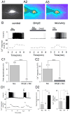
Figure 3. Nicotine increases intracellular Ca2+ concentrations in oriens/alveus interneurons via activation of non-α7 nAChRs
(A1–A3) Visualization of the fluorescent signal in oriens/alveus interneurons loaded with the Ca2+ indicator dye calcium green-1 through the recording pipette. (A1) A visualized oriens/alveus interneuron after dye loading. (A2, A3) Pseudo-color fluorescence imaging of a current-clamped oriens/alveus interneuron in the absence (A2) and presence (A3) of 10 μM nicotine. Note that application of nicotine produced detectable increases in fluorescent intensity at the soma. (B) Simultaneous recordings of electrical activity (Vm) and changes in Ca2+ fluorescence signal in a current-clamped oriens/alveus interneuron. Nicotine (10 μM; Nic)-induced changes in Vm and Ca2+ fluorescence signal were recorded in the absence (left) and presence of DHβE (2 μM; center) and 10 min after washout of DHβE (right). Recordings were carried out in the presence of DNQX (20 μM) and AP5 (40 μM). (C1) Summary graph showing the magnitude of depolarization observed in the presence of nicotine (10 μM) and nicotine (10 μM) + DHβE (2 μM). (C2) Summary graph showing Ca2+ fluorescence signal observed in the presence of nicotine (10 μM) and nicotine (10 μM) + DHβE (2 μM). (D1) Kainate (1 μM; KA)- and K+ (30 mM)-induced changes in Vm and Ca2+ fluorescence signal were simultaneously recorded. (D2) Summary graphs showing the magnitude of depolarization (left) and Ca2+ fluorescence signal (right) elicited by bath application of nicotine (10 μM), kainate (1 μM), and K+ (30 mM). *P < 0.05, ***P < 0.001.
Figure 4
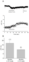
Figure 4. Nicotine elevates intracellular Ca2+ levels in oriens/alveus interneurons via Ca2+ entry through non-α7 nAChRs
(A) Simultaneous recordings of nicotine-induced currents (top) and changes in Ca2+ fluorescence signal (bottom) in a voltage-clamped oriens/alveus interneuron (at −70 mV). Recordings were carried out in the presence of DNQX (20 μM) and AP5 (40 μM). Nicotine (10 μM) was bath-applied as indicated by the horizontal bar. (B) Summary graph showing Ca2+ fluorescence signal (mean ± SEM) elicited in current-clamped (CC) and voltage-clamped (VC) oriens/alveus interneurons by bath application of nicotine (10 μM).
Figure 5
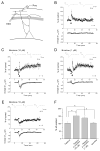
Figure 5. Nicotine promotes the induction of LTP at excitatory synapses onto oriens/alveus interneurons
(A) Scheme of recording setup showing the position of stimulating and recording electrodes. Oriens/alveus interneurons were voltage-clamped at −70 mV, and evoked EPSCs were recorded in the presence of AP5 (50 μM), bicuculline (10 μM), MLA (50 nM), and atropine (1 μM). (B–E) Changes in the amplitude of EPSCs were plotted as the percent change of initial baseline responses against time. Each trace above the graph in (B–E) was recorded at the time indicated. (B) A tetanus (100 pulses at 100Hz) alone failed to induce LTP. (C) A tetanus applied in the presence of nicotine (10 μM) induced LTP. (D) A tetanus applied in the presence of nicotine (1 μM) induced LTP. (E) Application of nicotine (10 μM) alone had no long-lasting effect on the amplitude of EPSCs. (C-E) Bath application of nicotine caused decreases in the amplitude of EPSCs (top) and increases in holding current (bottom). Administration of nicotine is indicated by the bar and delivery of tetanic stimulation is indicated by the arrow. (F) Histograms show the percent change (mean ± SEM) in the amplitude of EPSCs measured 50–60 min after delivery of tetanic stimulation in the absence and presence of nicotine, or after application of nicotine alone. *P < 0.05.
Figure 6
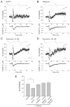
Figure 6. Ca2+ entry through non-α7 nAChRs facilitates LTP induction
Oriens/alveus interneurons were voltage-clamped at −70 mV, and evoked EPSCs were recorded in the presence of AP5 (50 μM), bicuculline (10 μM), MLA (50 nM), and atropine (1 μM). (A–D) Changes in the amplitude of EPSCs were plotted as the percent change of initial baseline responses against time. Each trace above the graph in (A–D) was recorded at the time indicated. (A) Nicotine-induced facilitation of LTP induction was blocked by intracellular perfusion of the calcium chelator BAPTA through the recording pipette (10 mM). (B) Nicotine still promoted LTP induction in the presence of nifedipine (30 μM). (C, D) Nicotine still facilitated LTP induction in the presence of 10 μM (C) and 100 μM (D) of ryanodine. (A–D) Bath application of nicotine caused decreases in the amplitude of EPSCs (top) and increases in holding current (bottom). Nifedipine (30 μM) or ryanodine (10 μM) was present throughout the entire recording period in (B) or (C), respectively. In the case of 100 μM ryanodine (D), the drug was present until 5 min after tetanus. Administration of nicotine is indicated by the bar and delivery of tetanic stimulation (100 pulses at 100Hz) is indicated by the arrow. (E) Histograms show the percent change (mean ± SEM) in the amplitude of EPSCs measured 50–60 min after delivery of tetanic stimulation, to compare the effects of different drugs on nicotine-induced facilitation of LTP induction. *P < 0.05.

Similar articles

Cited by

References

    1. Abdulla FA, Bradbury E, Calaminici MR, Lippiello PM, Wonnacott S, Gray JA, Sinden JD. Relationship between up-regulation of nicotine binding sites in rat brain and delayed cognitive enhancement observed after chronic or acute nicotinic receptor stimulation. Psychopharmacology. 1996;124:323–331. - PubMed
    1. Alkondon M, Pereira EF, Almeida LE, Randall WR, Albuquerque EX. Nicotine at concentrations found in cigarette smokers activates and desensitizes nicotinic acetylcholine receptors in CA1 interneurons of rat hippocampus. Neuropharmacology. 2000;39:2726–2739. - PubMed
    1. Alkondon M, Albuquerque EX. Nicotinic acetylcholine receptor alpha7 and alpha4beta2 subtypes differentially control GABAergic input to CA1 neurons in rat hippocampus. J Neurophysiol. 2001;86:3043–55. - PubMed
    1. Alkondon M, Albuquerque EX. Nicotinic receptor subtypes in rat hippocampal slices are differentially sensitive to desensitization and early in vivo functional up-regulation by nicotine and to block by bupropion. J Pharmacol Exp Ther. 2005;313:740–750. - PubMed
    1. Benowitz NL, Porchet H, Jacob P. Nicotine dependence and tolerance in man: pharmacokinetic and pharmacodynamic investigations. Prog Brain Res. 1989;79:279–287. - PubMed

Publication types

MeSH terms