. 2010 Jun;130(2):202-16.
doi: 10.1111/j.1365-2567.2009.03224.x. Epub 2010 Jan 27.

Differential regulation of toll-like receptor-2, toll-like receptor-4, CD16 and human leucocyte antigen-DR on peripheral blood monocytes during mild and severe dengue fever

Affiliations

Differential regulation of toll-like receptor-2, toll-like receptor-4, CD16 and human leucocyte antigen-DR on peripheral blood monocytes during mild and severe dengue fever

Elzinandes L Azeredo et al. Immunology. 2010 Jun.

Abstract

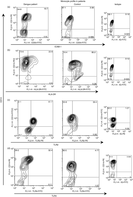
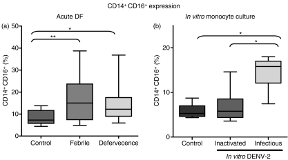
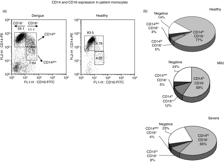
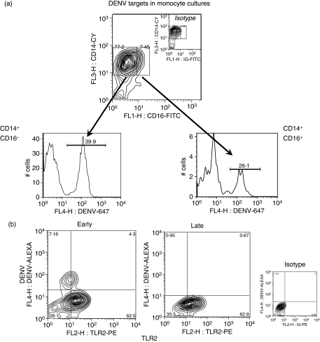
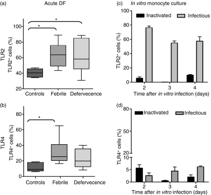
Dengue fever (DF), a public health problem in tropical countries, may present severe clinical manifestations as result of increased vascular permeability and coagulation disorders. Dengue virus (DENV), detected in peripheral monocytes during acute disease and in in vitro infection, leads to cytokine production, indicating that virus-target cell interactions are relevant to pathogenesis. Here we investigated the in vitro and in vivo activation of human peripheral monocytes after DENV infection. The numbers of CD14(+) monocytes expressing the adhesion molecule intercellular adhesion molecule 1 (ICAM-1) were significantly increased during acute DF. A reduced number of CD14(+) human leucocyte antigen (HLA)-DR(+) monocytes was observed in patients with severe dengue when compared to those with mild dengue and controls; CD14(+) monocytes expressing toll-like receptor (TLR)2 and TLR4 were increased in peripheral blood from dengue patients with mild disease, but in vitro DENV-2 infection up-regulated only TLR2. Increased numbers of CD14(+) CD16(+) activated monocytes were found after in vitro and in vivo DENV-2 infection. The CD14(high) CD16(+) monocyte subset was significantly expanded in mild dengue, but not in severe dengue. Increased plasma levels of tumour necrosis factor-alpha (TNF-alpha), interferon-gamma (IFN-gamma) and interleukin (IL)-18 in dengue patients were inversely associated with CD14(high) CD16(+), indicating that these cells might be involved in controlling exacerbated inflammatory responses, probably by IL-10 production. We showed here, for the first time, phenotypic changes on peripheral monocytes that were characteristic of cell activation. A sequential monocyte-activation model is proposed in which DENV infection triggers TLR2/4 expression and inflammatory cytokine production, leading eventually to haemorrhagic manifestations, thrombocytopenia, coagulation disorders, plasmatic leakage and shock development, but may also produce factors that act in order to control both intense immunoactivation and virus replication.


Figures

Figure 3
Figure 3
(a) Cellular flow cytometry profile showing size and granulosity of cells during dengue fever. (b) Peripheral blood mononuclear cells (PBMCs) were tri-labelled with specific monoclonal antibodies (mAbs) CD14, CD11c and human leucocyte antigen (HLA)-DR and analysed by flow cytometry within the monocyte gate. (c) Representative histograms are shown of HLA-DR expression among CD14+ CD11c+ monocytes between groups of patients with mild dengue, severe dengue and healthy individuals. Cy, cycrome; FITC, fluorescein isothiocyanate; PE, phycoerythrin.
Figure 1
Figure 1
Intercellular adhesion molecule 1 (ICAM-1) and human leucocyte antigen (HLA)-DR detected in monocytes during dengue fever (DF). Peripheral blood mononuclear cells (PBMCs) from healthy subjects or samples from dengue patients taken at the febrile phase or defervecence were analyzed using double-colour flow cytometry for (a) adhesion molecule ICAM-1 (CD54) (Controls, n = 10; Febrile, n = 15; Defervecence, n = 16) or (b) HLA-DR (Controls, n = 10; Febrile, n = 26; Defervecence, n = 24) expression on CD14+ cells. Box-and-whisker plot. The box extends from the 25th percentile to the 75th percentile and the line at the middle is the median. The error bars, or whiskers, extend down to the lowest value and up to the highest. Statistical significance was assessed using the Kruskal–Wallis test followed by Dunn’s Multiple Comparison Test. *P < 0·05 and **P < 0·01.
Figure 2
Figure 2
Flow cytometry profile of monocytes during dengue fever. Representative contour plots from patients during the acute phase of the disease and in healthy subjects and respective control isotypes. Peripheral blood mononuclear cells (PBMCs) were labelled as described in the Materials and methods and were analysed by flow cytometry within the monocyte gate. Ex vivo analysis of CD54, human leucocyte antigen (HLA)-DR, toll-like receptor (TLR)2 and TLR4 expression on CD14+ cells. The numbers in quadrants indicate the percentage of cells corresponding to the cell subset. ICAM-1, intercellular adhesion molecule 1.
Figure 5
Figure 5
CD14+ CD16+ expression on monocytes during acute dengue fever (DF) and in vitro monocyte culture. Box-and-whisker plots represent data from (a) peripheral blood mononuclear cells (PBMCs) from healthy subjects (n = 14) or samples from dengue patients taken at the febrile phase (n = 25) or during defervecence (n = 25) and analyzed using double-colour flow cytometry for CD16 expression in CD14+ cells. Statistical significance was assessed using the Kruskal–Wallis test followed by Dunn’s multiple comparison test. (b) Human monocytes were obtained from buffy coats and incubated with cell culture medium, or with heat-inactivated or infectious dengue virus 2 (DENV-2) (strain 16681). Box-and-whisker plots represent experiments performed with cells from seven different PBMC donors. Statistical significance was assessed using one-way analysis of variance (anova) followed by Tukey’s multiple comparison test. *P < 0·05, **P < 0·01.
Figure 6
Figure 6
Distribution of CD14 and CD16 on monocyte subsets in peripheral blood mononuclear cells (PBMCs) obtained from patients during acute dengue fever. Cells labelled with monoclonal antibodies (mAbs) against CD14 and CD16 can be separated into three distinct subsets: CD14high CD16; CD14high CD16+; and CD14low CD16+. (a) Scatter plots from a representative patient with dengue fever (DF) and a healthy individual were analyzed by flow cytometry within the monocyte gate. (b) Monocyte subset distribution during dengue fever. There was a significantly higher level of CD14high CD16+ cells in patients with mild dengue (12 ± 7%, n = 30) than in patients with severe dengue (8 ± 3%, n = 23) or in healthy subjects (4·5 ± 1·2%, n = 14). One-way analysis of variance (anova) followed by Tukey’s multiple comparison test with P < 0·05 and P < 0·001, respectively.
Figure 7
Figure 7
Dengue virus targets in monocyte cultures. Monocyte-enriched human peripheral blood mononuclear cells (PBMCs) (2 × 10⁵ cells/well) were incubated for 2 days with dengue virus 2 (DENV-2) (strain 16681). (a) Representative contour plot for CD14+ CD16+ expression during in vitro dengue infection triple-labelled with anti-CD14-cycrome (Cy), CD16-fluorescein isothiocyanate (FITC) and anti-DENV-Alexa 647 or their control antibodies (monocyte-gated cells). Representative histograms showing typical DENV-antigen (Ag) positive cells were detected on CD14+ CD16 classical monocytes and on CD14+ CD16+ monocytes by flow cytometry. Inactivated virus showed no detectable labelling (data not shown). (b) Toll-like receptor (TLR)2 and DENV-Ag expression in human monocytes. Monocytes, double labelled with anti-DENV-Alexa 647 and anti-TLR2-phycoerythrin (PE) (monocyte-gated cells), after 2 days (early) or 5 days (late) of infection with DENV-2 (16681). Quadrants were set by isotype-control antibodies.
Figure 4
Figure 4
Toll-like receptor (TLR)2 and TLR4 detected on monocytes from patients with dengue fever (DF) or after in vitro infection. Box-and-whisker plots represent data from TLR2 and TLR4 expression. (a, b) Peripheral blood mononuclear cells (PBMCs) from healthy subjects (n = 6) or samples from dengue patients taken on the fever day (n = 10) and during defervescence (n = 13). (c, d) Monocyte-enriched human PBMCs (2 × 10⁵ cells/well) were cultured for 2–4 days with DENV-2 (16681) or inactivated virus. Cultures were labelled with anti-TLR2 phycoerythrin (PE) or TLR-4 fluorescein isothiocyanate (FITC) conjugates, or their control antibodies. Statistical significance was assessed using the Kruskal–Wallis test followed by Dunn’s Multiple Comparison Test. *P < 0·05.

Similar articles

Cited by

References

    1. Nogueira RMR, de Araújo JMG, Schatzmayr HG. Dengue viruses in Brazil, 1986–2006. Revista Panamericana de Salud Pública/Pan. Am J Public Health. 2007;22:358–63. - PubMed
    1. Brazilian-Health-Ministry Secretaria de Vigilância em Saúde. Boletim epidemiológico, Secretaria de Vigilância em Saúde. Informe Epidemiológico da Dengue. Janeiro a Novembro de 2008. 2009. http://portal.saude.gov.br/portal/arquivos/pdf/boletim_dengue_janeiro_no... [accessed on 26 December 2009]
    1. Gibbons RV, Vaughn DW. Dengue: an escalating problem. BMJ. 2002;324:1563–6. - PMC - PubMed
    1. Avila-Aguero ML, Avila-Aguero CR, Um SL, Soriano-Fallas A, Canas-Coto A, Yan SB. Systemic host inflammatory and coagulation response in the dengue virus primo-infection. Cytokine. 2004;27:173–9. - PubMed
    1. Clyde K, Kyle JL, Harris E. Recent advances in deciphering viral and host determinants of dengue virus replication and pathogenesis. J Virol. 2006;80:11418–31. - PMC - PubMed

Publication types

MeSH terms