. 2010 Jan 29:11:8.
doi: 10.1186/1471-2091-11-8.

Orthophosphate binding at the dimer interface of Corynebacterium callunae starch phosphorylase: mutational analysis of its role for activity and stability of the enzyme

Affiliations

Orthophosphate binding at the dimer interface of Corynebacterium callunae starch phosphorylase: mutational analysis of its role for activity and stability of the enzyme

Mario Mueller et al. BMC Biochem. .

Abstract

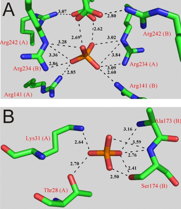
Background: Orthophosphate recognition at allosteric binding sites is a key feature for the regulation of enzyme activity in mammalian glycogen phosphorylases. Protein residues co-ordinating orthophosphate in three binding sites distributed across the dimer interface of a non-regulated bacterial starch phosphorylase (from Corynebacterium callunae) were individually replaced by Ala to interrogate their unknown function for activity and stability of this enzyme.

Results: While the mutations affected neither content of pyridoxal 5'-phosphate cofactor nor specific activity in phosphorylase preparations as isolated, they disrupted (Thr28-->Ala, Arg141-->Ala) or decreased (Lys31-->Ala, Ser174-->Ala) the unusually strong protective effect of orthophosphate (10 or 100 mM) against inactivation at 45 degrees C and subunit dissociation enforced by imidazole, as compared to wild-type enzyme. Loss of stability in the mutated phosphorylases appeared to be largely due to weakened affinity for orthophosphate binding. Binding of sulphate mimicking the crystallographically observed "non-covalent phosphorylation" of the phosphorylase at the dimer interface did not have an allosteric effect on the enzyme activity.

Conclusions: The phosphate sites at the subunit-subunit interface of C. callunae starch phosphorylase appear to be cooperatively functional in conferring extra kinetic stability to the native dimer structure of the active enzyme. The molecular strategy exploited for quaternary structure stabilization is to our knowledge novel among dimeric proteins. It can be distinguished clearly from the co-solute effect of orthophosphate on protein thermostability resulting from (relatively weak) interactions of the ligand with protein surface residues.


Figures

Figure 1
Figure 1
Close-up structures of the orthophosphate binding sites at the dimer interface of CcGlgP. Panels A (Tower helix region; P-site) and B (CAP region) are drawn using the x-ray structure of CcGlgP (PDB-accession code 2C4M). There are two identical CAP sites at the subunit-subunit interface, related by the internal symmetry of the enzyme dimer. H-bond distances are in Å, and coordinating residues contributed from subunit A or B are indicated in brackets.
Figure 2
Figure 2
Inactivation of R141A and S174A at 45°C. A 50 mM triethanolamine buffer, pH 7.0, was used. The protein concentration was 35 μg/ml (A) and 20 μg/ml (B). Symbols show the experimental data and solid lines are the corresponding straight-line fits. Representative data are shown. Conditions: no Pi added (full circles); 10 mM Pi (open circles).
Figure 3
Figure 3
Dissociation of enzyme subunits in R141A enforced by imidazole. R141A as isolated is a mixture consisting of the native dimer and a small amount of a tetrameric form that is also active. The N-terminal His-tag causes the tetramerization [15]. The absorbance traces are in arbitrary units (a.u.). Elution profiles are shown for R141A prior to (dashed line) and after incubation in the presence of 0.4 M imidazole for 30 min (dotted line), 60 min (solid line) and 140 min (dashed-dotted line). The observed peaks correspond to tetrameric (t; 362 kDa), dimeric (d; 181 kDa) and monomeric (m; 90.6 kDa) forms of the protein. A high-molecular mass peak is also visible in some traces, presumably showing soluble aggregated protein. Loss of phosphorylase activity in the presence of imidazole is correlated with the extent to which monomer formation had occurred (data not shown). Note that all samples contained the same protein concentration (0.4 mg/ml) prior to the incubation with imidazole. The decrease in peak area for the eluted protein forms as the incubation time in the presence of imidazole increased probably reflects loss of protein due to aggregation. Insoluble aggregates are removed by centrifugation prior to gel filtration. The protein concentration of the sample applied to the Superdex column was not measured.
Figure 4
Figure 4
Enhancement by sulphate of enzyme activity in the synthesis direction for wild-type and mutated forms of CcGlgP. A: Effect of sulphate on Vs for the wild-type phosphorylase. (full circles), 25 nM protein, 50 mM G1P; (full triangles) 25 nM protein, 5.0 mM G1P; (open circles), 50 nM protein, 50 mM G1P. Starch (20 g/l) was the acceptor substrate. B: Specific activities (grey bars; left y-axis) for wild-type and mutated phosphorylases determined at a protein concentration of 2.5 nM in the absence of (NH4)2SO4 using 50 mM G1P and 20 g/l maltodextrin as substrates. Relative enhancement of the respective activity by 10 mM (NH4)2SO4 is shown with black bars (right y-axis). All results are representative data.

Similar articles

Cited by

References

    1. Schinzel R, Nidetzky B. Bacterial α-glucan phosphorylases. FEMS Microbiol Lett. 1999;171:73–79. - PubMed
    1. Coutinho PM, Deleury E, Davies GJ, Henrissat B. An evolving hierarchical family classification for glycosyltransferases. J Mol Biol. 2003;328:307–317. doi: 10.1016/S0022-2836(03)00307-3. - DOI - PubMed
    1. Palm D, Klein HW, Schinzel R, Buehner M, Helmreich EJ. The role of pyridoxal 5'-phosphate in glycogen phosphorylase catalysis. Biochemistry. 1990;29:1099–1107. doi: 10.1021/bi00457a001. - DOI - PubMed
    1. Withers SG, Madsen NB, Sprang SR, Fletterick RJ. Catalytic site of glycogen phosphorylase: structural changes during activation and mechanistic implications. Biochemistry. 1982;21:5372–5382. doi: 10.1021/bi00264a039. - DOI - PubMed
    1. Johnson LN. Glycogen phosphorylase: control by phosphorylation and allosteric effectors. FASEB J. 1992;6:2274–2282. - PubMed

Publication types

LinkOut - more resources