Anti-viral state segregates two molecular phenotypes of pancreatic adenocarcinoma: potential relevance for adenoviral gene therapy
- PMID: 20113473
- PMCID: PMC2845551
- DOI: 10.1186/1479-5876-8-10
Anti-viral state segregates two molecular phenotypes of pancreatic adenocarcinoma: potential relevance for adenoviral gene therapy
Abstract
Background: Pancreatic ductal adenocarcinoma (PDAC) remains a leading cause of cancer mortality for which novel gene therapy approaches relying on tumor-tropic adenoviruses are being tested.
Methods: We obtained the global transcriptional profiling of primary PDAC using RNA from eight xenografted primary PDAC, three primary PDAC bulk tissues, three chronic pancreatitis and three normal pancreatic tissues. The Affymetrix GeneChip HG-U133A was used. The results of the expression profiles were validated applying immunohistochemical and western blot analysis on a set of 34 primary PDAC and 10 established PDAC cell lines. Permissivity to viral vectors used for gene therapy, Adenovirus 5 and Adeno-Associated Viruses 5 and 6, was assessed on PDAC cell lines.
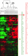
Results: The analysis of the expression profiles allowed the identification of two clearly distinguishable phenotypes according to the expression of interferon-stimulated genes. The two phenotypes could be readily recognized by immunohistochemical detection of the Myxovirus-resistance A protein, whose expression reflects the activation of interferon dependent pathways. The two molecular phenotypes discovered in primary carcinomas were also observed among established pancreatic adenocarcinoma cell lines, suggesting that these phenotypes are an intrinsic characteristic of cancer cells independent of their interaction with the host's microenvironment. The two pancreatic cancer phenotypes are characterized by different permissivity to viral vectors used for gene therapy, as cell lines expressing interferon stimulated genes resisted to Adenovirus 5 mediated lysis in vitro. Similar results were observed when cells were transduced with Adeno-Associated Viruses 5 and 6.
Conclusion: Our study identified two molecular phenotypes of pancreatic cancer, characterized by a differential expression of interferon-stimulated genes and easily recognized by the expression of the Myxovirus-resistance A protein. We suggest that the detection of these two phenotypes might help the selection of patients enrolled in virally-mediated gene therapy trials.
Figures


Similar articles
-
Efficient Gene Delivery and Expression in Pancreas and Pancreatic Tumors by Capsid-Optimized AAV8 Vectors.Hum Gene Ther Methods. 2017 Feb;28(1):49-59. doi: 10.1089/hgtb.2016.089. Hum Gene Ther Methods. 2017. PMID: 28125909 Free PMC article.
-
Oncolytic Adenovirus Expressing ST13 Increases Antitumor Effect of Tumor Necrosis Factor-Related Apoptosis-Inducing Ligand Against Pancreatic Ductal Adenocarcinoma.Hum Gene Ther. 2020 Aug;31(15-16):891-903. doi: 10.1089/hum.2020.024. Epub 2020 Jun 30. Hum Gene Ther. 2020. PMID: 32475172
-
Delivery of interferon alpha using a novel Cox2-controlled adenovirus for pancreatic cancer therapy.Surgery. 2012 Jul;152(1):114-22. doi: 10.1016/j.surg.2012.02.017. Epub 2012 Apr 11. Surgery. 2012. PMID: 22503318 Free PMC article.
-
Development of gene therapy to target pancreatic cancer.Cancer Sci. 2004 Apr;95(4):283-9. doi: 10.1111/j.1349-7006.2004.tb03204.x. Cancer Sci. 2004. PMID: 15072584 Free PMC article. Review.
-
[Organoids from pancreatic ductal adenocarcinoma].Med Sci (Paris). 2020 Jan;36(1):57-62. doi: 10.1051/medsci/2019259. Epub 2020 Feb 4. Med Sci (Paris). 2020. PMID: 32014099 Review. French.
Cited by
-
Immunotherapeutic Challenges for Pediatric Cancers.Mol Ther Oncolytics. 2019 Aug 28;15:38-48. doi: 10.1016/j.omto.2019.08.005. eCollection 2019 Dec 20. Mol Ther Oncolytics. 2019. PMID: 31650024 Free PMC article. Review.
-
Targeting Palbociclib-Resistant Estrogen Receptor-Positive Breast Cancer Cells via Oncolytic Virotherapy.Cancers (Basel). 2019 May 16;11(5):684. doi: 10.3390/cancers11050684. Cancers (Basel). 2019. PMID: 31100952 Free PMC article.
-
Breaking resistance of pancreatic cancer cells to an attenuated vesicular stomatitis virus through a novel activity of IKK inhibitor TPCA-1.Virology. 2015 Nov;485:340-54. doi: 10.1016/j.virol.2015.08.003. Epub 2015 Aug 29. Virology. 2015. PMID: 26331681 Free PMC article.
-
Tumor Restrictions to Oncolytic Virus.Biomedicines. 2014 Apr 17;2(2):163-194. doi: 10.3390/biomedicines2020163. Biomedicines. 2014. PMID: 28548066 Free PMC article. Review.
-
Expanding the Spectrum of Pancreatic Cancers Responsive to Vesicular Stomatitis Virus-Based Oncolytic Virotherapy: Challenges and Solutions.Cancers (Basel). 2021 Mar 9;13(5):1171. doi: 10.3390/cancers13051171. Cancers (Basel). 2021. PMID: 33803211 Free PMC article. Review.
References
Publication types
MeSH terms
Substances
LinkOut - more resources
Full Text Sources
Other Literature Sources
Medical

