

Expression of epithelial calcium transport system in rat cochlea and vestibular labyrinth
- PMID: 20113508
- PMCID: PMC2825184
- DOI: 10.1186/1472-6793-10-1
Expression of epithelial calcium transport system in rat cochlea and vestibular labyrinth
Abstract
Background: The low luminal Ca2+ concentration of mammalian endolymph in the inner ear is required for normal hearing and balance. We recently reported the expression of mRNA for a Ca2+-absorptive transport system in primary cultures of semicircular canal duct (SCCD) epithelium.
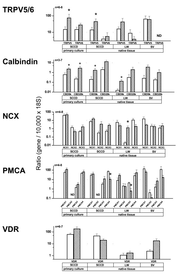
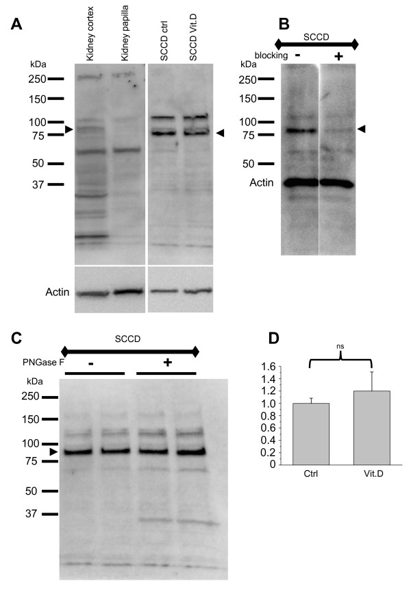
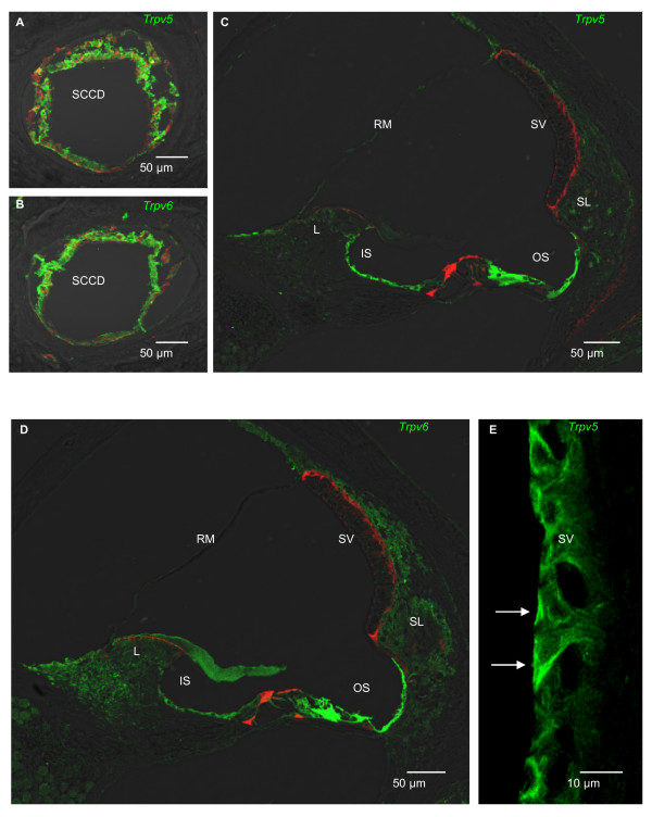
Results: We now identify this system in native vestibular and cochlear tissues by qRT-PCR, immunoblots and confocal immunolocalization. Transcripts were found and quantified for several isoforms of epithelial calcium channels (TRPV5, TRPV6), calcium buffer proteins (calbindin-D9K, calbindin-D28K), sodium-calcium exchangers (NCX1, NCX2, NCX3) and plasma membrane Ca2+-ATPase (PMCA1, PMCA2, PMCA3, and PMCA4) in native SCCD, cochlear lateral wall (LW) and stria vascularis (SV) of adult rat as well as Ca2+ channels in neonatal SCCD. All components were expressed except TRPV6 in SV and PMCA2 in SCCD. 1,25-(OH)2vitamin D3 (VitD) significantly up-regulated transcripts of TRPV5 in SCCD, calbindin-D9K in SCCD and LW, NCX2 in LW, while PMCA4 in SCCD and PMCA3 in LW were down-regulated. The expression of TRPV5 relative to TRPV6 was in the sequence SV > Neonatal SCCD > Adult SCCD > LW > primary culture SCCD. Expression of TRPV5 protein from primary culture of SCCD did not increase significantly when cells were incubated with VitD (1.2 times control; P > 0.05). Immunolocalization showed the distribution of TRPV5 and TRPV6. TRPV5 was found near the apical membrane of strial marginal cells and both TRPV5 and TRPV6 in outer and inner sulcus cells of the cochlea and in the SCCD of the vestibular system.
Conclusions: These findings demonstrate for the first time the expression of a complete Ca2+ absorptive system in native cochlear and vestibular tissues. Regulation by vitamin D remains equivocal since the results support the regulation of this system at the transcript level but evidence for control of the TRPV5 channel protein was lacking.
Figures
Similar articles
-
Vitamin D upregulates expression of ECaC1 mRNA in semicircular canal.Biochem Biophys Res Commun. 2005 Jun 17;331(4):1353-7. doi: 10.1016/j.bbrc.2005.04.053. Biochem Biophys Res Commun. 2005. PMID: 15883024
-
Dietary calcium and 1,25-dihydroxyvitamin D3 regulate transcription of calcium transporter genes in calbindin-D9k knockout mice.J Reprod Dev. 2009 Apr;55(2):137-42. doi: 10.1262/jrd.20139. Epub 2008 Dec 24. J Reprod Dev. 2009. PMID: 19106481
-
The expression and implication of TRPV5, Calbindin-D28k and NCX1 in idiopathic hypercalciuria.J Huazhong Univ Sci Technolog Med Sci. 2008 Oct;28(5):580-3. doi: 10.1007/s11596-008-0520-z. Epub 2008 Oct 10. J Huazhong Univ Sci Technolog Med Sci. 2008. PMID: 18846343
-
Coordinated control of renal Ca2+ handling.Kidney Int. 2006 Feb;69(4):650-4. doi: 10.1038/sj.ki.5000169. Kidney Int. 2006. PMID: 16518325 Review.
-
On the molecular mechanism of intestinal calcium transport.Adv Exp Med Biol. 1989;249:45-65. doi: 10.1007/978-1-4684-9111-1_5. Adv Exp Med Biol. 1989. PMID: 2543194 Review.
Cited by
-
Osteoporosis increases the risk of benign paroxysmal positional vertigo: a nested case-control study using a national sample cohort.Eur Arch Otorhinolaryngol. 2019 Feb;276(2):335-342. doi: 10.1007/s00405-018-5230-y. Epub 2018 Dec 3. Eur Arch Otorhinolaryngol. 2019. PMID: 30511104
-
Association between vitamin D, vitamin D supplementation and benign paroxysmal positional vertigo: a systematic review and meta-analysis.Front Neurol. 2025 Apr 16;16:1560616. doi: 10.3389/fneur.2025.1560616. eCollection 2025. Front Neurol. 2025. PMID: 40308226 Free PMC article.
-
Prevention of Recurrent Benign Paroxysmal Positional Vertigo: The Role of Combined Supplementation with Vitamin D and Antioxidants.Audiol Res. 2022 Aug 22;12(4):445-456. doi: 10.3390/audiolres12040045. Audiol Res. 2022. PMID: 36004953 Free PMC article.
-
Clinical Outcomes in Patients With Benign Paroxysmal Positional Vertigo and Vitamin D Deficiency: A Singaporean Perspective.Cureus. 2024 May 15;16(5):e60325. doi: 10.7759/cureus.60325. eCollection 2024 May. Cureus. 2024. PMID: 38883121 Free PMC article.
-
Effect of the serum 25-hydroxyvitamin D level on risk for short-term residual dizziness after successful repositioning in benign paroxysmal positional vertigo stratified by sex and onset age.Front Neurol. 2023 Mar 31;14:1144958. doi: 10.3389/fneur.2023.1144958. eCollection 2023. Front Neurol. 2023. PMID: 37064183 Free PMC article.
References
-
- Marcus DC, Wangemann P. In: Physiology and Pathology of Chloride Transporters and Channels in the Nervous System--From molecules to diseases. Alvarez-Leefmans FJ, Delpire E, editor. Elsevier; 2009. Cochlear and Vestibular Function and Dysfunction; pp. 421–433.
-
- Peng JB, Brown EM, Hediger MA. Apical entry channels in calcium-transporting epithelia. News Physiol Sci. 2003;18:158–163. - PubMed
Publication types
MeSH terms
Substances
Grants and funding
LinkOut - more resources
Full Text Sources
Molecular Biology Databases
Miscellaneous
