

MicroRNA-21 inhibitor sensitizes human glioblastoma cells U251 (PTEN-mutant) and LN229 (PTEN-wild type) to taxol
- PMID: 20113523
- PMCID: PMC2824710
- DOI: 10.1186/1471-2407-10-27
MicroRNA-21 inhibitor sensitizes human glioblastoma cells U251 (PTEN-mutant) and LN229 (PTEN-wild type) to taxol
Abstract
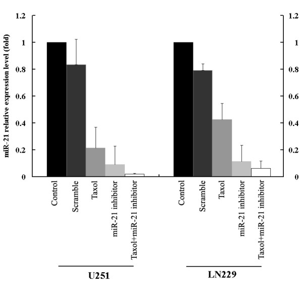
Background: Substantial data indicate that the oncogene microRNA 21 (miR-21) is significantly elevated in glioblastoma multiforme (GBM) and regulates multiple genes associated with cancer cell proliferation, apoptosis, and invasiveness. Thus, miR-21 can theoretically become a target to enhance the chemotherapeutic effect in cancer therapy. So far, the effect of downregulating miR-21 to enhance the chemotherapeutic effect to taxol has not been studied in human GBM.
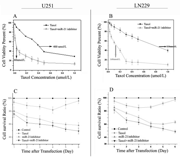
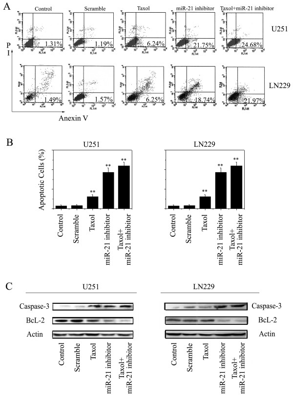
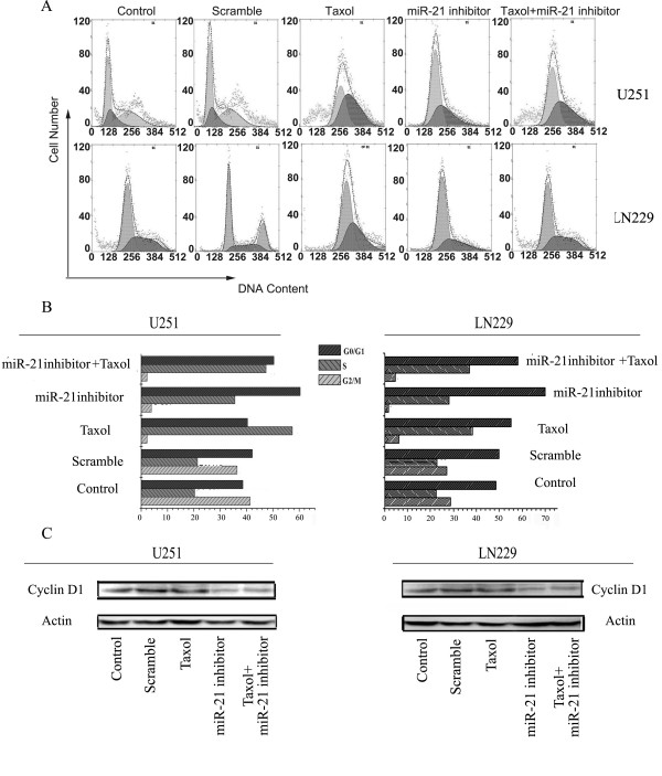
Methods: Human glioblastoma U251 (PTEN-mutant) and LN229 (PTEN wild-type) cells were treated with taxol and the miR-21 inhibitor (in a poly (amidoamine) (PAMAM) dendrimer), alone or in combination. The 50% inhibitory concentration and cell viability were determined by the MTT assay. The mechanism between the miR-21 inhibitor and the anticancer drug taxol was analyzed using the Zheng-Jun Jin method. Annexin V/PI staining was performed, and apoptosis and the cell cycle were evaluated by flow cytometry analysis. Expression of miR-21 was investigated by RT-PCR, and western blotting was performed to evaluate malignancy related protein alteration.
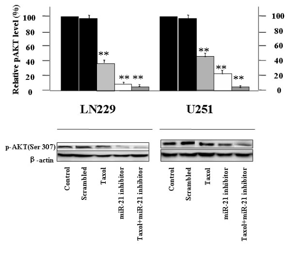
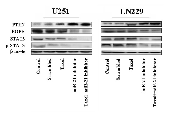
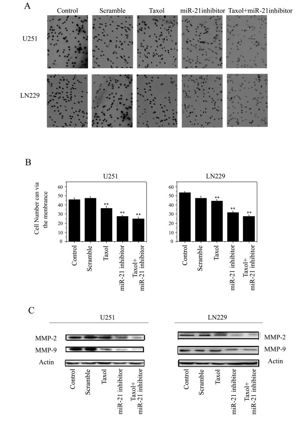
Results: IC(50) values were dramatically decreased in cells treated with miR-21 inhibitor combine with taxol, to a greater extent than those treated with taxol alone. Furthermore, the miR-21 inhibitor significantly enhanced apoptosis in both U251 cells and LN229 cells, and cell invasiveness was obviously weakened. Interestingly, the above data suggested that in both the PTEN mutant and the wild-type GBM cells, miR-21 blockage increased the chemosensitivity to taxol. It is worth noting that the miR-21 inhibitor additively interacted with taxol on U251cells and synergistically on LN229 cells. Thus, the miR-21 inhibitor might interrupt the activity of EGFR pathways, independently of PTEN status. Meanwhile, the expression of STAT3 and p-STAT3 decreased to relatively low levels after miR-21 inhibitor and taxol treatment. The data strongly suggested that a regulatory loop between miR-21 and STAT3 might provide an insight into the mechanism of modulating EGFR/STAT3 signaling.
Conclusions: Taken together, the miR-21 inhibitor could enhance the chemo-sensitivity of human glioblastoma cells to taxol. A combination of miR-21 inhibitor and taxol could be an effective therapeutic strategy for controlling the growth of GBM by inhibiting STAT3 expression and phosphorylation.
Figures
Similar articles
-
Anti-miR-155 oligonucleotide enhances chemosensitivity of U251 cell to taxol by inducing apoptosis.Cell Biol Int. 2012 Jul;36(7):653-9. doi: 10.1042/CBI20100918. Cell Biol Int. 2012. PMID: 22276743
-
MicroRNA-424-5p enhances chemosensitivity of breast cancer cells to Taxol and regulates cell cycle, apoptosis, and proliferation.Mol Biol Rep. 2021 Feb;48(2):1345-1357. doi: 10.1007/s11033-021-06193-4. Epub 2021 Feb 8. Mol Biol Rep. 2021. PMID: 33555529
-
Co-inhibition of microRNA-10b and microRNA-21 exerts synergistic inhibition on the proliferation and invasion of human glioma cells.Int J Oncol. 2012 Sep;41(3):1005-12. doi: 10.3892/ijo.2012.1542. Epub 2012 Jul 3. Int J Oncol. 2012. PMID: 22766763
-
Downregulation of miR-21 inhibits EGFR pathway and suppresses the growth of human glioblastoma cells independent of PTEN status.Lab Invest. 2010 Feb;90(2):144-55. doi: 10.1038/labinvest.2009.126. Epub 2010 Jan 4. Lab Invest. 2010. PMID: 20048743
-
STAT3 regulation of glioblastoma pathogenesis.Curr Mol Med. 2009 Jun;9(5):580-90. doi: 10.2174/156652409788488739. Curr Mol Med. 2009. PMID: 19601808 Free PMC article. Review.
Cited by
-
Stratifying risk of recurrence in stage II colorectal cancer using deregulated stromal and epithelial microRNAs.Oncotarget. 2015 Mar 30;6(9):7262-79. doi: 10.18632/oncotarget.3225. Oncotarget. 2015. PMID: 25788261 Free PMC article.
-
MicroRNAs delivery into human cells grown on 3D-printed PLA scaffolds coated with a novel fluorescent PAMAM dendrimer for biomedical applications.Sci Rep. 2018 Sep 17;8(1):13888. doi: 10.1038/s41598-018-32258-9. Sci Rep. 2018. PMID: 30224665 Free PMC article.
-
Correlation of MicroRNA 132 Up-regulation with an Unfavorable Clinical Outcome in Patients with Primary Glioblastoma Multiforme Treated with Radiotherapy Plus Concomitant and Adjuvant Temozolomide Chemotherapy.Transl Oncol. 2013 Dec 1;6(6):742-8. doi: 10.1593/tlo.13553. eCollection 2013 Dec 1. Transl Oncol. 2013. PMID: 24466377 Free PMC article.
-
Effects of miR-21 on proliferation and apoptosis of WT cells via PTEN/Akt pathway.Exp Ther Med. 2020 Mar;19(3):2155-2160. doi: 10.3892/etm.2019.8376. Epub 2019 Dec 27. Exp Ther Med. 2020. PMID: 32104279 Free PMC article.
-
Role of 3'-untranslated region translational control in cancer development, diagnostics and treatment.World J Biol Chem. 2014 Feb 26;5(1):40-57. doi: 10.4331/wjbc.v5.i1.40. World J Biol Chem. 2014. PMID: 24600513 Free PMC article. Review.
References
-
- Davies MA, Lu Y, Sano T. Adenoviral transgene expression of MMAC/PTEN in human glioma cells inhibits Akt activation and induces anoikis. Cancer Res. 1998;58:5285–90. - PubMed
Publication types
MeSH terms
Substances
LinkOut - more resources
Full Text Sources
Other Literature Sources
Medical
Research Materials
Miscellaneous
