

Characterization and expression of a heart-selective alternatively spliced variant of alpha II-spectrin, cardi+, during development in the rat
- PMID: 20114050
- PMCID: PMC3537504
- DOI: 10.1016/j.yjmcc.2010.01.001
Characterization and expression of a heart-selective alternatively spliced variant of alpha II-spectrin, cardi+, during development in the rat
Abstract
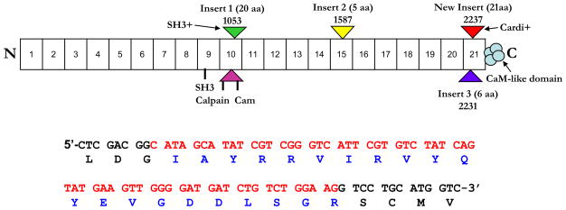
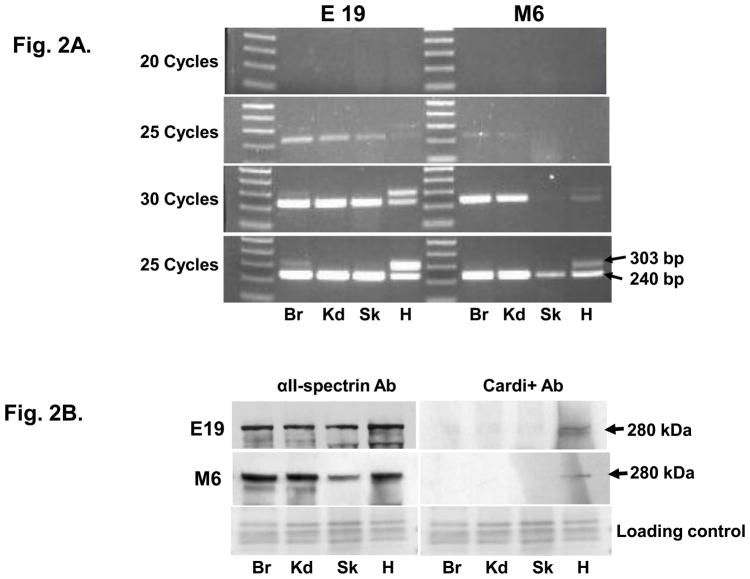
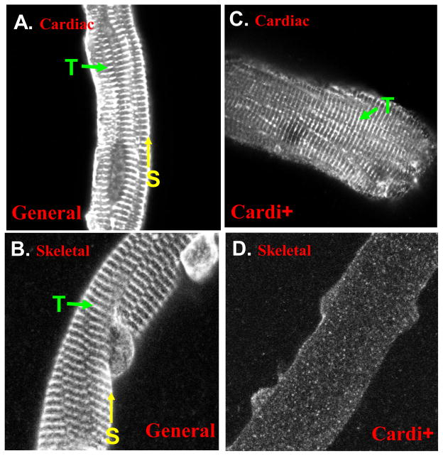
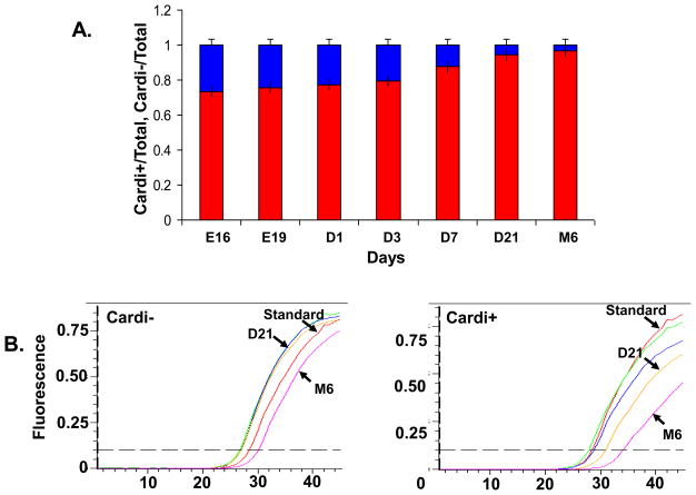
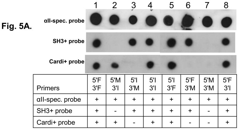
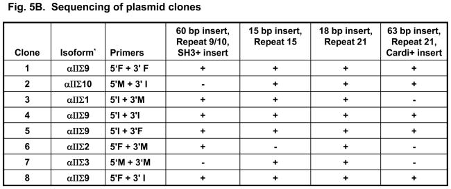
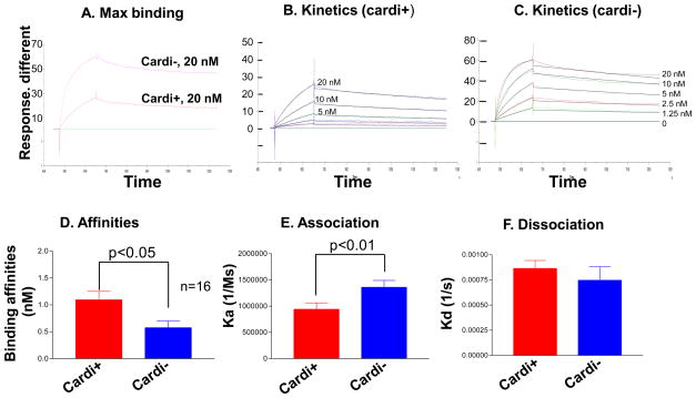
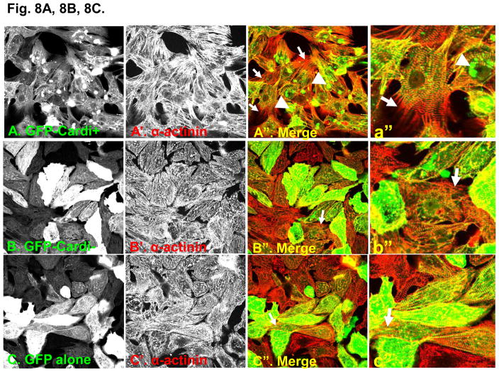
Spectrin is a large, flexible protein that stabilizes membranes and organizes proteins and lipids into microdomains in intracellular organelles and at the plasma membrane. Alternative splicing occurs in spectrins, but it is not yet clear if these small variations in structure alter spectrin's functions. Three alternative splice sites have been identified previously for alpha II-spectrin. Here we describe a new alternative splice site, a 21-amino acid sequence in the 21st spectrin repeat that is only expressed in significant amounts in cardiac muscle (GenBank GQ502182). The insert, which we term alpha II-cardi+, results in an insertion within the high affinity nucleation site for binding of alpha-spectrins to beta-spectrins. To assess the developmental regulation of the alpha II-cardi+ isoform, we used qRT-PCR and quantitative immunoblotting methods to measure the levels of this form and the alpha II-cardi- form in the cardiac muscles of rats, from embryonic day 16 (E16) through adulthood. The alpha II-cardi+ isoform constituted approximately 26% of the total alpha II-spectrin in E16 hearts but decreased to approximately 6% of the total after 3 weeks of age. We used long-range RT-PCR and Southern blot hybridization to examine possible linkage of the alpha II-cardi+ alternatively spliced sequence with alternatively spliced sequences of alpha II-spectrin that had been previously reported. We identified two new isoforms of alpha II-spectrin containing the cardi+ insert. These were named alpha II Sigma 9 and alpha II Sigma 10 in accordance with the spectrin naming conventions. In vitro studies of recombinant alpha II-spectrin polypeptides representing the two splice variants of alpha II-spectrin, alpha II-cardi+ and alpha II-cardi-, revealed that the alpha II-cardi+ subunit has lower affinity for the complementary site in repeats 1-4 of betaII-spectrin, with a K(D) value of approximately 1 nM, as measured by surface plasmon resonance (SPR). In addition, the alpha II-cardi+ form showed 1.8-fold lower levels of binding to its site on beta II-spectrin than the alpha II-cardi- form, both by SPR and blot overlay. This suggests that the 21-amino acid insert prevented some of the alpha II-cardi+ form from interacting with beta II-spectrin. Fusion proteins expressing the alpha II-cardi+ sequence within the two terminal spectrin repeats of alpha II-spectrin were insoluble in solution and aggregated in neonatal myocytes, consistent with the possibility that this insert removes a significant portion of the protein from the population that can bind beta subunits. Neonatal rat cardiomyocytes infected with adenovirus encoding GFP-fusion proteins of repeats 18-21 of alpha II-spectrin with the cardi+ insert formed many new processes. These processes were only rarely seen in myocytes expressing the fusion protein lacking the insert or in controls expressing only GFP. Our results suggest that the embryonic mammalian heart expresses a significant amount of alpha II-spectrin with a reduced avidity for beta-spectrin and the ability to promote myocyte growth.
(c) 2010 Elsevier Ltd. All rights reserved.
Figures


Comment in
-
Cardiac spectrins: alternative splicing encodes functional diversity.J Mol Cell Cardiol. 2010 Jun;48(6):1031-2. doi: 10.1016/j.yjmcc.2010.02.002. Epub 2010 Feb 6. J Mol Cell Cardiol. 2010. PMID: 20144617 Free PMC article. No abstract available.
Similar articles
-
Brain and muscle express a unique alternative transcript of alphaII spectrin.Biochemistry. 1999 Nov 30;38(48):15721-30. doi: 10.1021/bi991458k. Biochemistry. 1999. PMID: 10625438
-
Beta II-spectrin (fodrin) and beta I epsilon 2-spectrin (muscle) contain NH2- and COOH-terminal membrane association domains (MAD1 and MAD2).J Biol Chem. 1994 Nov 18;269(46):29212-9. J Biol Chem. 1994. PMID: 7961888
-
A partial structural repeat forms the heterodimer self-association site of all beta-spectrins.J Biol Chem. 1994 Apr 15;269(15):11400-8. J Biol Chem. 1994. PMID: 8157672
-
The role of βII spectrin in cardiac health and disease.Life Sci. 2018 Jan 1;192:278-285. doi: 10.1016/j.lfs.2017.11.009. Epub 2017 Nov 9. Life Sci. 2018. PMID: 29128512 Free PMC article. Review.
-
Evolution of spectrin function in cytoskeletal and membrane networks.Biochem Soc Trans. 2009 Aug;37(Pt 4):796-803. doi: 10.1042/BST0370796. Biochem Soc Trans. 2009. PMID: 19614597 Review.
Cited by
-
Mechanistic Insights of Phenobarbital-Mediated Activation of Human but Not Mouse Pregnane X Receptor.Mol Pharmacol. 2019 Sep;96(3):345-354. doi: 10.1124/mol.119.116616. Epub 2019 Jul 10. Mol Pharmacol. 2019. PMID: 31436536 Free PMC article.
-
Spectrin-based skeleton as an actor in cell signaling.Cell Mol Life Sci. 2012 Jan;69(2):191-201. doi: 10.1007/s00018-011-0804-5. Epub 2011 Aug 30. Cell Mol Life Sci. 2012. PMID: 21877118 Free PMC article. Review.
-
Supporting the heart: Functions of the cardiomyocyte's non-sarcomeric cytoskeleton.J Mol Cell Cardiol. 2019 Jun;131:187-196. doi: 10.1016/j.yjmcc.2019.04.002. Epub 2019 Apr 9. J Mol Cell Cardiol. 2019. PMID: 30978342 Free PMC article. Review.
-
Defining new mechanistic roles for αII spectrin in cardiac function.J Biol Chem. 2019 Jun 14;294(24):9576-9591. doi: 10.1074/jbc.RA119.007714. Epub 2019 May 7. J Biol Chem. 2019. PMID: 31064843 Free PMC article.
-
Cardiac spectrins: alternative splicing encodes functional diversity.J Mol Cell Cardiol. 2010 Jun;48(6):1031-2. doi: 10.1016/j.yjmcc.2010.02.002. Epub 2010 Feb 6. J Mol Cell Cardiol. 2010. PMID: 20144617 Free PMC article. No abstract available.
References
-
- Cianci CD, Zhang Z, Pradhan D, Morrow JS. Brain and muscle express a unique alternative transcript of alphaII spectrin. Biochemistry. 1999;38:15721–30. - PubMed
-
- Dubreuil RR. Functional links between membrane transport and the spectrin cytoskeleton. J Membr Biol. 2006;211:151–61. - PubMed
-
- Bennett PM, Baines AJ, Lecomte MC, Maggs AM, Pinder JC. Not just a plasma membrane protein: in cardiac muscle cells alpha-II spectrin also shows a close association with myofibrils. J Muscle Res Cell Motil. 2004;25:119–26. - PubMed
-
- Baines AJ, Pinder JC. The spectrin-associated cytoskeleton in mammalian heart. Front Biosci. 2005;10:3020–33. - PubMed
Publication types
MeSH terms
Substances
Grants and funding
LinkOut - more resources
Full Text Sources
