The effect of NELL1 and bone morphogenetic protein-2 on calvarial bone regeneration
- PMID: 20116699
- PMCID: PMC3113462
- DOI: 10.1016/j.joms.2009.03.066
The effect of NELL1 and bone morphogenetic protein-2 on calvarial bone regeneration
Abstract
Purpose: Most craniofacial birth defects contain skeletal components that require bone grafting. Although many growth factors have shown potential for use in bone regeneration, bone morphogenetic proteins (BMPs) are the most osteoinductive. However, supraphysiologic doses, high cost, and potential adverse effects stimulate clinicians and researchers to identify complementary molecules that allow a reduction in dose of BMP-2. Because NELL1 plays a key role as a regulator of craniofacial skeletal morphogenesis, especially in committed chondrogenic and osteogenic differentiation, and a previous synergistic mechanism has been identified, NELL1 is an ideal molecule for combination with BMP-2 in calvarial defect regeneration. We investigated the effect of NELL1 and BMP-2 on bone regeneration in vivo.
Materials and methods: BMP-2 doses of 589 and 1,178 ng were grafted into 5-mm critical-sized rat calvarial defects, as compared with 589 ng of NELL1 plus 589 ng of BMP-2 and 1,178 ng of NELL1 plus 1,178 ng of BMP-2, and bone regeneration was analyzed.
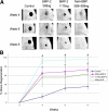
Results: Live micro-computed tomography data showed increased bone formation throughout 4 to 8 weeks in all groups but a significant improvement when the lower doses of each molecule were combined. High-resolution micro-computed tomography and histology showed more mature and complete defect healing when the combination of NELL1 plus BMP-2 was compared with BMP-2 alone at lower doses.
Conclusion: The observed potential synergy has significant value in the future treatment of patients with craniofacial defects requiring extensive bone grafting that would normally entail extraoral autogenous bone grafts or doses of BMP-2 in milligrams.
Copyright 2010 American Association of Oral and Maxillofacial Surgeons. Published by Elsevier Inc. All rights reserved.
Figures


References
-
- Bardach J, Morris H. Multidisciplinary Management of Cleft Lip and Palate. Saunders; Philadelphia: 1990.
-
- Cohen M, MacLean R. Craniosynostosis: Diagnosis, Evaluation, and Management. Oxford; New York: 2000.
-
- Cohen MM., Jr Craniofacial disorders caused by mutations in homeobox genes MSX1 and MSX2. J Craniofac Genet Dev Biol. 2000;20:19. - PubMed
-
- Canady JW, Zeitler DP, Thompson SA, et al. Suitability of the iliac crest as a site for harvest of autogenous bone grafts. Cleft Palate Craniofac J. 1993;30:579. - PubMed
-
- Franceschi RT. Biological approaches to bone regeneration by gene therapy. J Dent Res. 2005;84:1093. - PubMed
Publication types
MeSH terms
Substances
Grants and funding
LinkOut - more resources
Full Text Sources
Other Literature Sources

