. 2010 Apr 10;399(2):221-30.
doi: 10.1016/j.virol.2010.01.010. Epub 2010 Feb 8.

Temperature-sensitive mutant in the vaccinia virus E6 protein produce virions that are transcriptionally inactive

Affiliations

Temperature-sensitive mutant in the vaccinia virus E6 protein produce virions that are transcriptionally inactive

Olga Boyd et al. Virology. .

Abstract

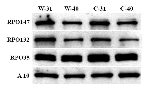
The vaccinia virus E6R gene encodes a late protein that is packaged into virion cores. A temperature-sensitive mutant was used to study the role of this protein in viral replicative cycle. Cts52 has a P226L missense mutation in the E6R gene, shows a two-log reduction in plaque formation, but displays normal patterns of gene expression, late protein processing and DNA replication during infection. Mutant virions produced at 40 degrees C were similar in their morphology to wt virions grown at 40 degrees C. The particle to infectivity ratio was 50 times higher in purified Cts52 grown at 40 degrees C when compared to the mutant grown at permissive temperature. In vitro characterization of Cts-52 particles grown at 40 degrees C revealed no differences in protein composition or in DNA content and the mutant virions could bind and enter cells. However, core particles prepared from Cts52 grown at 40 degrees C failed to transcribe in vitro. Our results show that E6 in the virion has either a direct or an indirect role in viral transcription.


Figures

Fig. 1
Fig. 1. One-step growth and DNA replication in cells infected with wt and mutants in the E6R gene
A) BSC40 cells were infected at moi = 10 pfu/cell with wt (VACV-WR) or Cts52 incubated at 31 or 40°C. At indicated times post-infection samples were removed and virus yields were determined by plaque titration at 31°C. B) BSC40 cells were infected wt (VACV-IHD-W), or Dts41or Dts80 as described above. Virus yield was determined by plaque assay at 31°C after 24 and 48 hpi. C) BSC-40 cells were infected wt (VACV-WR) or Cts52, incubated at 31 or 40 °C as described above. At indicated times post-infection, cell extracts were prepared and applied in triplicate on nylon membranes, and hybridization to a 32P labeled vaccinia virus DNA probe was performed as described under Methods. The numbers (arbitrary units) express the average value of three different samples. wt-31°C (◆), wt-40 °C (∎), Cts52-31 °C (▴), Cts52-40 °C (●).
Fig. 2
Fig. 2. Virus protein synthesis, processing of p4a, and accumulation of E6
BSC40 cells were infected at moi = 10 pfu/cell, with wt or Cts52 and incubated at 31 or 40 °C. A) At different times post-infection, the cells were pulse-labeled with 35S methionine, cells lysates were prepared, the proteins separated on SDS-PAGE, and gels were dried and autoradiographed. The virus used in the infection and the hpi of the pulse label is indicated at the top of each column. The incubation temperature is indicated to the left of the figure and the approximate molecular weights, in kDa, are indicated to the right of each autoradiogram. B-C) At indicated times after infection, cells lysates were prepared, the proteins separated by SDS-PAGE, transferred to a nitrocellulose membrane and probe against viral proteins: p4a /A10 (B), or E6 (C). The virus, the temperature of incubation, and the hpi are indicated on the top of the figure, the estimated molecular weights in kDa are indicated on the left, and the arrow on the right represents the relevant proteins.
Fig. 3
Fig. 3. Electron micrographs of wt and mutants-infected cells
BSC40 cells were infected with wt or mutant virus at a moi = 10 pfu/cell, incubated at 40 °C for 24 (A-C) and 48 hs (D -F) and processed for electron microscopy as described in Methods. (A) wt; (B - D) Cts52; (E) Dts41; (F) Dts80. IV= immature virions; MV= mature virions; C= crescents; N= nucleus; F= DNA factories.
Fig. 4
Fig. 4. Analysis of the DNA content and protein composition of purified wt and mutant particles
A) Samples of purified wt and Cts52 were diluted and the amount of virions determined by A260nm and indicated on the top of each lane were applied onto a nylon membrane, processed and hybridized to 32P VACV DNA fragment. B) The blot was analyzed with a phosphor-imager apparatus, the data were normalized and graphed as described in Methods. C) The polypeptide composition of purified virions was analyzed by SDS-PAGE and Coomassie blue staining. The amount of virions determined by A260nm is indicated on the top of each lane and the approximate molecular weight marker is indicated on the left of the gel. D) Partition of purified virions into core and membrane fraction and Western blot analysis. 0.06 A260 nm units (~0.7 μg) of each virion sample was resuspended in core buffer and processed as described under Methods. The gene product corresponding to each antiserum used with the predicted molecular weight is indicated to the left of each row, the virus sample probed is indicated on the top of the figure. C= core fraction, M = membrane fraction. Wt31, wt grown at 31°C; Wt-40, wt grown at 40 °C; Cts- 31, Cts52 grown at 31°C; Cts40, Cts52 grown at 40 °C.
Fig. 5
Fig. 5. Analysis of the binding and entry process of wt and mutant virions
BSC40 cells were infected with moi= 1000 particles of wt (A, C, E, G) or Cts52 (B, D, F, H) virus grown at 31 °C (A, B, E, F) or 40 °C (C, D, G, H). The cells were processed and stained for viral membrane protein A27 (green) (A-D) or viral core protein A4 (red) (E-H), and DNA was stained with Dapi (blue). The cells were analyzed by confocal microscopy as described in Methods.
Fig 6
Fig 6. RNA synthesis directed by purified wt and mutants particles
A) Total core RNA synthesis. Purified virions grown at different temperature were permeabilized with NP40 and DTT treatment, incubated in a reaction mixture that contains [α32P] CTP, unlabelled GTP, ATP and UTP, MgCl2 as described in Methods. At indicated times, samples were removed, and the acid precipitable radioactivity was determined in a liquid scintillation counter. wt-31°C, (◆); wt-40°C, (∎); Cts52-31 °C, (●); Cts52-40 °C, (▴). B) RNA extrusion from cores. Purified virions grown at 31 °C were activated for RNA as described above. At indicated times samples were removed and the core and the soluble fraction were separated by centrifugation as described in Methods. wt-core, (∎); wt-supernatant, (◆); Cts52-core (▴); Cts-52-supernantant, (●).
Fig 7
Fig 7. Western blot analysis of the RNA polymerases subunits in wt and mutants particles
0 .06 A260 nm units (~0.7 μg) of each virion sample was resuspended in SDS-sample buffer and processed as described under Methods. The gene product corresponding to each antiserum used is indicated to the left of each row, the virus sample probed is indicated on the top of the figure. The core protein A10 was used as a gel loading control.

References

    1. Afonso CL, Tulman ER, Lu Z, Oma E, Kutish GF, Rock DL. The genome of Melanoplus sanguinipes entomopoxvirus. J.Virol. 1999;73:533–552. - PMC - PubMed
    1. Ausubel FM, Brent R, Kingston RE, Moore DD, Seidman JG, Smith JA, Struhl K. Current protocols in molecular biology. John Wiley & Sons; New York: 1994.
    1. Bablanian R, Baxt B, Sonnabend JA, Esteban M. Studies on the mechanisms of vaccinia virus cytopathic effects. II. Early cell rounding is associated with virus polypeptide synthesis. J.Gen.Virol. 1978;39:403–413. - PubMed
    1. Boyd O, Turner P, Moyer RW, Condit RC, Moussatche N. Involvement of E6 protein in vaccinia virus morphogenesis. Virology. 2010 - PMC - PubMed
    1. Byrd CM, Hruby DE. Vaccinia virus proteolysis--a review. Rev.Med.Virol. 2006;16:187–202. - PMC - PubMed

Publication types

MeSH terms