Review
. 2010 Aug;58(4):258-66.
doi: 10.1016/j.patbio.2009.11.001. Epub 2010 Feb 8.

Pathogenesis of hepatitis B virus infection

Affiliations
Review

Pathogenesis of hepatitis B virus infection

F V Chisari et al. Pathol Biol (Paris). 2010 Aug.

Abstract

The adaptive immune response is thought to be responsible for viral clearance and disease pathogenesis during hepatitis B virus infection. It is generally acknowledged that the humoral antibody response contributes to the clearance of circulating virus particles and the prevention of viral spread within the host while the cellular immune response eliminates infected cells. The T cell response to the hepatitis B virus (HBV) is vigorous, polyclonal and multispecific in acutely infected patients who successfully clear the virus and relatively weak and narrowly focussed in chronically infected patients, suggesting that clearance of HBV is T cell dependent. The pathogenetic and antiviral potential of the cytotoxic T lymphocyte (CTL) response to HBV has been proven by the induction of a severe necroinflammatory liver disease following the adoptive transfer of HBsAg specific CTL into HBV transgenic mice. Remarkably, the CTLs also purge HBV replicative intermediates from the liver by secreting type 1 inflammatory cytokines thereby limiting virus spread to uninfected cells and reducing the degree of immunopathology required to terminate the infection. Persistent HBV infection is characterized by a weak adaptive immune response, thought to be due to inefficient CD4+ T cell priming early in the infection and subsequent development of a quantitatively and qualitatively ineffective CD8+ T cell response. Other factors that could contribute to viral persistence are immunological tolerance, mutational epitope inactivation, T cell receptor antagonism, incomplete down-regulation of viral replication and infection of immunologically privileged tissues. However, these pathways become apparent only in the setting of an ineffective immune response, which is, therefore, the fundamental underlying cause. Persistent infection is characterized by chronic liver cell injury, regeneration, inflammation, widespread DNA damage and insertional deregulation of cellular growth control genes, which, collectively, lead to cirrhosis of the liver and hepatocellular carcinoma.


Figures

Figure 1
Figure 1
Liver gene expression profile during HBV and HCV infection [9]. (A) Genes correlated with viremia in all acutely HBV infected chimpanzees. No genes correlated positively or negatively with the level of intrahepatic HBV DNA during the time course of acute HBV infection in all three animals. (B) Intrahepatic gene expression correlated with viremia in three HCV infected chimpanzees [107]. (C) Liver gene expression profile associated with viral clearance in all three acutely HBV-infected chimpanzees and (D) that associated with clearance in HCV infected chimpanzees [107]. Gene identities in HBV and HCV infected animals are described in Wieland et al [9] and Su et al [107], respectively. Blue and green lines show the intrahepatic HBV DNA or serum HCV RNA as a percentage (% max) of the corresponding peak levels, respectively. For optimal visualization, gene expression levels (red lines) are shown after normalization to the 10th and 90th percentiles for each gene. Values on the x axis represent weeks after inoculation with HBV. Adapted from Wieland et al. (2004) PNAS vol. 101 pp. 6669–74 and Su et al. (2002) PNAS vol. 99 pp. 11181–6.
Figure 2
Figure 2
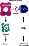
Noncytopathic clearance of HBV from the hepatocyte by T cell- derived cytokines. On antigen recognition, CD8-positive CTL deliver an apoptotic signal to their target cells, killing them. They also secrete IFNγ and TNF-α, cytokines that abolish viral replication and HBV gene expression in vivo, potentially curing them. Reprinted/amended from Am J Pathol 2000, 156:1117–1132 with permission from the American Society for Investigative Pathology.
Figure 3
Figure 3
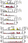
Peripheral CD4+ T cell responses against HBV core protein and intrahepatic CD8+ T cell responses in chimpanzees infected with a dose range of HBV as described in Asabe et al. [21]. The upper panel in each figure represents the serum HBV DNA as a black line and sALT as a yellow shaded area and the results of peripheral CD4+ T cell ELISPOT assays are overlaid as black bars. Cryopreserved PBMCs were thawed and stimulated in vitro with HBV core protein and the numbers of IFNγ producing cells were determined by ELISPOT assay as described [21]. The data are shown as number of spots at each time point minus the number of spots before inoculation per million PBMCs. The lower panel shows the total number of intrahepatic HBV-specific CD8+ T cells per 102 total CD8+ T cells as filled blue bars (right axis) and fold induction of intrahepatic CD8 mRNA compared to two preinoculation time points as a shaded red area (left axis). Intrahepatic lymphocytes were expanded antigen-nonspecifically in vitro and tested with all the corresponding Patr/peptide multimer complexes as described [21]. nt: not tested. *: tested and negative. Reproduced/amended with permission from American Society for Microbiology from Journal of Virology, 2009, vol 83, pp. 9652–62, DOI: 10.1128/JVI.00867-09.
Figure 4
Figure 4
Course of HBV infections, peripheral CD4+ T cell responses against HBV core protein, and intrahepatic CD8+ T cell responses in chimpanzees with or without CD4 immunodepletion as described in Asabe et al [21]. Serum HBV DNA levels are shown as a black line and sALT as a yellow shaded area. Horizontal bars represent serum HBe and HBs antigen levels and the open horizontal bars represent the presence of anti-HBc, anti-HBe, and anti-HBs antibodies. The amount of each protein is reflected by the thickness of each bar as indicated in the legend. The numbers of CD4+ T cells per μL of whole blood were shown as closed squares (top panel, right axis). Arrows on the top panels represent injections of control antibody (a) or anti-CD4 antibody (b). Peripheral CD4+ T cell IFNγ ELISPOT assays against HBV core protein (second panel) and detection of intrahepatic HBV-specific CD8+ T cells (bottom panel, left axis) were as described in Figure 3 except that freshly prepared cells were used instead of using cryopreserved cells. Fold induction of intrahepatic CD8 mRNA compared to two preinoculation time points is shown as a shaded red area (bottom panel, right axis). *: tested and negative. Reproduced/amended with permission from American Society for Microbiology from Journal of Virology, 2009, vol 83, pp. 9652–62, DOI: 10.1128/JVI.00867-09.
Figure 5
Figure 5
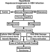
The chronic injury → HCC hypothesis. According to this hypothesis, a vigorous (+++) immune response to HBV leads to viral clearance while an absent (−) immune response leads to the “healthy” carrier state, and an intermediate (+) immune response produces chronic hepatitis. This indolent necroinflammatory liver disease is characterized by chronic liver cell necrosis which stimulates a sustained regenerative response. The inflammatory component includes activated macrophages that are a rich source of free radicals. The collaboration of these mitogenic and mutagenic stimuli has the potential to cause cellular and viral DNA damage, chromosomal abnormalities, genetic mutations, etc, that deregulate cellular growth control in a multistep process that eventually leads to hepatocellular carcinoma. Reprinted from Am J Pathol 2000, 156:1117–1132 with permission from the American Society for Investigative Pathology.

Similar articles

Cited by

References

    1. Chisari FV. Rous-Whipple Award Lecture. Viruses, immunity, and cancer: lessons from hepatitis B. Am J Pathol. 2000;156:1117–32. - PMC - PubMed
    1. Guidotti LG, Chisari FV. Immunobiology and pathogenesis of viral hepatitis. Annu Rev Pathol. 2006;1:23–61. - PubMed
    1. Guidotti LG, Matzke B, Schaller H, Chisari FV. High-level hepatitis B virus replication in transgenic mice. J Virol. 1995;69:6158–69. - PMC - PubMed
    1. Guidotti LG, Rochford R, Chung J, Shapiro M, Purcell R, Chisari FV. Viral clearance without destruction of infected cells during acute HBV infection. Science. 1999;284:825–9. - PubMed
    1. Thimme R, Wieland S, Steiger C, Ghrayeb J, Reimann KA, Purcell RH, et al. CD8(+) T cells mediate viral clearance and disease pathogenesis during acute hepatitis B virus infection. J Virol. 2003;77:68–76. - PMC - PubMed

Publication types

MeSH terms