Review
. 2010 Apr;13(2):133-41.
doi: 10.1016/j.mib.2009.12.013. Epub 2010 Jan 29.

Protein histidine kinases: assembly of active sites and their regulation in signaling pathways

Affiliations
Review

Protein histidine kinases: assembly of active sites and their regulation in signaling pathways

Richard C Stewart. Curr Opin Microbiol. 2010 Apr.

Abstract

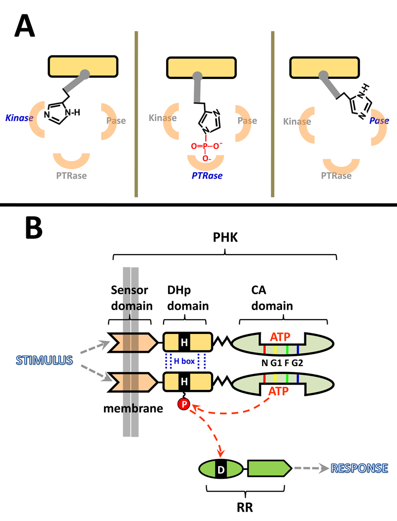
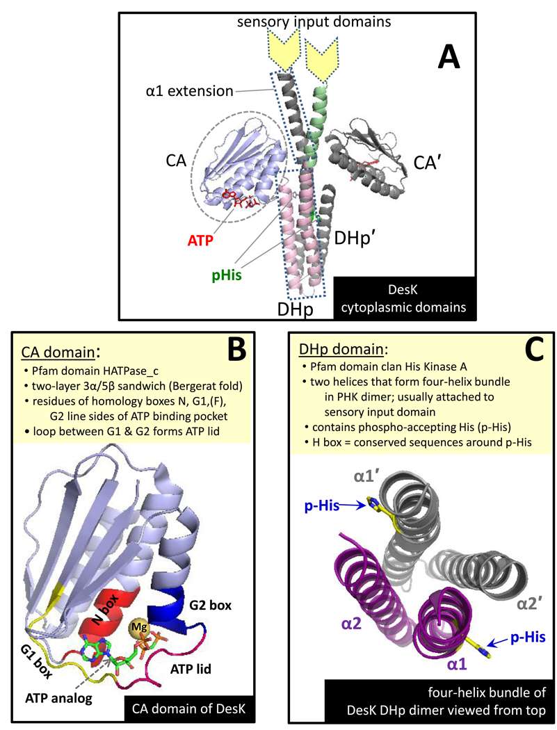
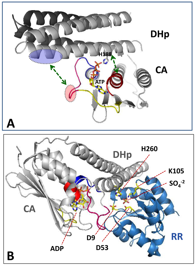
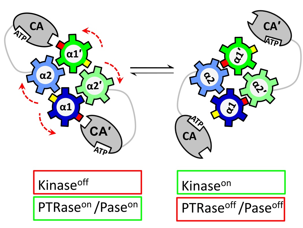
Protein histidine kinases (PHKs) function in Two Component Signaling pathways utilized extensively by bacteria and archaea. Many PHKs participate in three distinct, but interrelated signaling reactions: autophoshorylation, phosphotransfer (to a partner Response Regulator (RR) protein), and dephosphorylation of this RR. Detailed biochemical and structural characterization of several PHKs has revealed how the domains of these proteins can interact to assemble the three active sites that promote the necessary chemistry and how these domain interactions might be regulated in response to sensory input: the relative orientation of helices in the PHK dimerization domain can reorient, via cogwheeling (rotation) and kinking (bending), to effect changes in PHK activities that probably involve sequestration/release of the PHK catalytic domain by the dimerization domain.


Figures

Figure 1
Figure 1
Schematic diagram of the role played by PHKs in two-component signal transduction systems (TCSs). (A) Many PHKs have three distinct but interrelated enzymatic activities that involve positioning the phospho-accepting His in three active sites. The active sites for phosphotransferase (PTRase) and phosphatase(Pase) activities are likely to be very similar but are portrayed as being physically distinct for the purpose of illustration. Although the diagram depicts the phospho-accepting His rotating from site to site, this reorientation might also involve movement of the active sites relative to an essentially stationary phospho-accepting His. (B) PHKs function as homodimers that autophosphorylate then pass their phosphoryl group to an aspartate side chain located in the receiver domain of a cognate response regulator protein. Each PHK monomer has three distinct structural/functional domains: a transmembrane sensor, a DHp domain, and a CA domain. Sequence comparisons have defined a homology box within DHp (H box) that spans the phospho-accepting histidine. In addition, there are homology boxes (5–10 amino acids) located within the CA domain at/near the ATP binding pocket: N, G1 (sometimes called the D box), and G2 (sometimes called the G box) are conserved in all PHKs, while the F box is present in some, but not all, PHKs (note that DesK portrayed in Fig. 2 and Fig. 3 lacks the lacks an F box) [9,61,62]. The diagram depicts autophosphorylation via an intradimer cis mechanism; some PHKs utilize a trans mechanism.
Figure 2
Figure 2
Summary of key features of PHK structure as illustrated with B. subtilis DesK. (A) The crystal structure of the butterfly-shaped dimer formed by a DesK fragment that includes the CA and DHp domains as well as a helical extension of α1 of the DHp. The protomer in back is colored grey. Color coding for the front protomer: CA domain (blue), DHp helices (pink), extension of DHp helix (green). This panel was modeled after Fig. 1 of Jacques et al. [55•] (B) The CA domain of DesK with nonhydrolyzable ATP analog ADPCP bound. The location of the conserved homology box residues are shown in different colors as well as the ‘ATP-lid’ (magenta loop that folds over the polyphosphate groups of ATP). Note that DesK does not have an F box [9]. (C) A top-down view of the four-helix bundle formed by the dimerized DHp domain (with CA domain and helical extensions removed for purposes of illustration). α1 and α2 of one protomer are purple; helices of the second protomer are grey. The short connector linking α1 to α2 is at the bottom of the helices from this perspective, and the membrane/sensor input side would be the top. This figure was created using coordinates from PDB ID 3GIE (DesKDHp+CA H188E mutant) manipulated in PyMol to replace the mutant side chain with the phospho-accepting His (H188).
Figure 3
Figure 3
Views of the PHK autokinase and phosphotransferase active sites. (A) A PHK poised for autophosphorylation: DesKDHp+CA with ATP bound. This figure was generated by manually docking the one DesK CA domain onto the four-helix bundle of a DHp dimer (as described by Albanesi et al. [38••] (docking by rigid body rotation about a pivot point in the hinge linking DHp to CA). Coordinates for this structure were kindly provided by Dr. Alejandro Buschiazzo and correspond to Fig. S4 in reference [38••]. This docking orients the phospho-accepting His near the γ –phosphoryl of bound ATP and allows some complementary interdomain electrostatic interactions (green arrows), for example between acidic side chains of the ATP-lid (red oval) and basic side chains of the DHp (blue oval). Color coding of homology boxes of the CA domain (N, G1, G2, and ATP-lid) is the same as in Fig. 2. (B) A PHK poised for phosphotransfer: HK853DHp+CA bound to RR468 (generated using PDB 3DGE). Two molecules of R468 bind to the dimeric HK853, but in this diagram only one molecule of each protein is shown to improve clarity (i.e., only two helices of the four-helix bundle are shown). The PHK:RR complex brings the phospho-accepting His (H260) of HK853 close to D53 of RR468 (the phosphorylation site of RR468) and close to RR side chains that catalyze phosphotransfer (e.g., D9, D10, M55, T83, and K105); a sulfate ion occupies a position that may mimic that of phosphate during phosphotransfer reactions. Two key RR side chains (M55 and K105) interact with the PHK (M55 with E348 in the CA domain; and K105 with R263 and T267 in the DHp domain); their abilities to contribute to catalysis of the phosphotransfer reaction might be influenced by these associations. However, RR468, like all response regulators, can catalyze its own phosphorylation using small molecule phosphodonors (such as acetyl phosphate) in the absence of any PHK [63], and so it is likely that, like other RRs, RR468 does the ‘heavy lifting’ in catalyzing the PHK→RR phosphotransfer reaction [64], while the PHK might make a comparatively small contribution by altering the positions or efficacy of the catalytic scaffold provided by the RR. The PHK might, in addition, contribute to the general hydrophobic environment in which this catalytic scaffold operates, an environment expected to enhance the strength of charge-charge and H-bonding interactions [36•,40].
Figure 4
Figure 4
Schematic diagram depicting how rearrangement of the helices within the DHp domain could alter PHK activities. The DHp helices of the four-helix bundle formed in the PHK dimer are depicted (top-down view) as cogwheels. In the starting structure (left), a surface (red cog) of the DHp is not buried within the bundle and is available to sequester the CA domain: CA cannot access the phospho-accepting His. In this conformation, the PHK is not active as an autokinase, but the DHp does have interaction surfaces for RR binding, so it can function as phosphotransferase (if it has been phosphorylated) or as a phosphatase (if it has not). Signaling events can cause cogwheel rotation of the helices (by 60° in this diagram to match that reported by Albanesi et al. [38••]). In this new orientation (right), the red cog of the DHp is no longer accessible, and the CA domain has been released: now it can access the DHp surface (yellow cog) to complete assembly of the kinase active site, including the phospho-accepting His: now the PHK is active as an autokinase. This reorientation also inhibits phosphotransferase and/or phosphatase activities of the PHK, either because it hides DHp surfaces important for RR binding or because the CA now competes effectively with the RR for binding surface on the DHp.

Similar articles

Cited by

References

    1. Hoch JA, Silhavy TJ. Two-Component Signal Transduction. Washington, DC: ASM Press; 1995.
    1. Galperin MY. Structural classification of bacterial response regulators: diversity of output domains and domain combinations. J Bacteriol. 2006;46:4169–4182. - PMC - PubMed
    1. Gao R, Stock AM. Biological Insights from Structures of Two-Component Proteins. Ann Rev Microbiol. 2009;63:133–154. Both PHK afficianados and newcomers will appreciate this well written and up-to-date review.

    1. Saito H. Histidine phosphorylation and two-component signaling in eukaryotic cells. Chem Rev. 2001;101:2497–2509. - PubMed
    1. Chauhan N, Calderone R. Two-Component Signal Transduction Proteins as Potential Drug Targets in Medically Important Fungi. Infection and Immunity. 2008;76:4795–4803. - PMC - PubMed

Publication types

MeSH terms