Review
. 2010 Jul;211(1):1-8.
doi: 10.1016/j.atherosclerosis.2009.12.027. Epub 2009 Dec 29.

The metabolism of triglyceride-rich lipoproteins revisited: new players, new insight

Affiliations
Review

The metabolism of triglyceride-rich lipoproteins revisited: new players, new insight

Geesje M Dallinga-Thie et al. Atherosclerosis. 2010 Jul.

Abstract

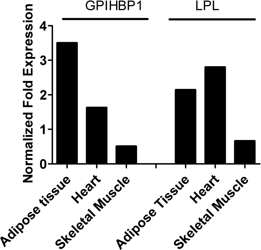
Peripheral lipoprotein lipase (LPL)-mediated lipolysis of triglycerides is the first step in chylomicron/VLDL clearance involving heparan sulfate proteoglycans (HSPGs) displayed at the cell surface of the capillaries in adipose tissue, heart and skeletal muscle. The newly generated chylomicron remnant particles are then cleared by the liver, whereas VLDL remnant particles are either further modified, through the action of hepatic lipase (HL) and cholesteryl ester transfer protein (CETP), into LDL particles or alternatively directly cleared by the liver. Two proteins, lipase maturation factor 1 (LMF1) and glycosylphosphatidylinositol-anchored high density lipoprotein binding protein 1 (GPIHBP1), have been recently identified and have revised our current understanding of LPL maturation and LPL-mediated lipolysis. Moreover, new insights have been gained with respect to hepatic remnant clearance using genetically modified mice targeting the sulfation of HSPGs and even deletion of the most abundant heparan sulfate proteoglycan: syndecan1. In this review, we will provide an overview of novel data on both peripheral TG hydrolysis and hepatic remnant clearance that will improve our knowledge of plasma triglyceride metabolism.


Figures

Figure 1
Figure 1. Protein Structure of LMF1
LMF1 contains 5 membrane-spanning regions, with its N-terminus located in the cytosol and the C-terminus within the ER lumen. The function of lipase maturation resides in a domain of unknown function (DUF1222, indicated by black line) that comprises about 65% of the C-terminus of LMF1. Partial truncations of this domain caused by cld, Y439X and W464X result in loss of a C-terminal portion of the domain located within the ER lumen, and result in impaired activity of LMF1 to support lipase maturation. The lipase interaction site and a glycosylation site at residue 430 are indicated
Figure 2
Figure 2. Tissue expression profile of GPIHBP1 and LPL
The expression profile of GPIHBP1 and LPL mRNA in human adipose tissue, heart and skeletal muscle (first choice human total RNA Survey panel, Ambion, Foster City, Ca, USA).[76] The mRNA was reversed transcribed and used for qPCR of GPIHBP1 and LPL. 36B4 is used as a house keeping gene.
Figure 3
Figure 3. A Model for GPIHBP1
Figure 3 A shows the consensus sequence of the Ly-6 motif.[77,78] The conserved cysteine residues are presented in yellow, whereas the non-conserved cysteine residues are shown in white. GPIHBP1 consists of 10 cysteine residues in the Ly-6 domain which will form 5 S-S bonds that determine its characteristic three-finger structure.[79] The consensus sequence of CD59 is very similar to GPIHBP1. The model has been build using the loop model procedure of MODELER 9v6. The model with the best DOPE (Discrete Optimized Protein Energy) score was selected. Based on this model we predict the structure of GPIHBP1 as shown in figure 3B. The cysteine residues are located in the global core of the protein. The three loops are formed based on the presence of 6 β-strands. Additionally the two putative glycosylation sites (Asn-76 and Asn-82) are shown. The p.Q115P is located adjacent to the 7th cysteine residue, whereas the p.C65Y mutation is the first cysteine residue of the Ly-6 domain. We hypothesize that both mutations may interfere with proper folding of the Ly-6 domain and consequently with the interaction between the acidic and the Ly-6 domain.
Figure 4
Figure 4. Peripheral TG Hydrolysis
LPL is synthesized in parenchymal cells, maturated by LMF1 and secreted in the subendothelial space where it binds to syndecan1. LPL will then be transcytosed through the endothelial cell to reach the cell surface at the luminal site where GPIHBP1 is present, probably in lipid rafts together with syndecan1 and fatty acid transporters such as CD36.[6,9] The generated remnant particle can then go to the liver. The fatty acids will be locally internalized by CD36.
Figure 5
Figure 5. Hepatic Remnant Clearance
View of hepatocytes with sinusoids and bile canaliculi. View of hepatoctes, with sinusoids and bile canuculi (green). Hepatic remnant clearance involves sequestration of the particle through the fenestrae. On its journey through the extracellular matrix the remnant particle will acquire additional apoE molecules which enables rapid binding to the LDL receptor or syndecan1 receptor, the most abundant HSPG in hepatocytes, which internalizes remnant particles.[72] LRP1 only facilitates uptake after modification of the particle by syndecan1. Both the LDLr and syndecan1 contribute equally to remnant clearance.

Similar articles

Cited by

References

    1. Nordestgaard BG, Benn M, Schnohr P, Tybjaerg-Hansen A. Nonfasting triglycerides and risk of myocaridal infarction, ischemic heart disease, and death in men and women. J. Am. Med. Ass. 2007;298:299–308. - PubMed
    1. Goldberg IJ, Merkel M. Lipoprotein lipase: physiology, biochemistry, and molecular biology. Front Biosci. 2001;6:D388–D405. - PubMed
    1. Bensadoun A. Lipoprotein lipase. Annu. Rev. Nutr. 1991;11:217–237. - PubMed
    1. Olivecrona T, Bengtsson-Olivecrona G. Lipoprotein lipase and hepatic lipase. Curr. Opin. Lipidol. 1993;4:187–196.
    1. Wang H, Eckel RH. Lipoprotein Lipase: from gene to obesity. Am. J. Physiol Endocrinol. Metab. 2009;297:E271–E288. - PubMed

Publication types