. 2010 Jul;148(1):87-95.
doi: 10.1016/j.surg.2009.12.004. Epub 2010 Feb 1.

Real-time intra-operative near-infrared fluorescence identification of the extrahepatic bile ducts using clinically available contrast agents

Affiliations

Real-time intra-operative near-infrared fluorescence identification of the extrahepatic bile ducts using clinically available contrast agents

Aya Matsui et al. Surgery. 2010 Jul.

Abstract

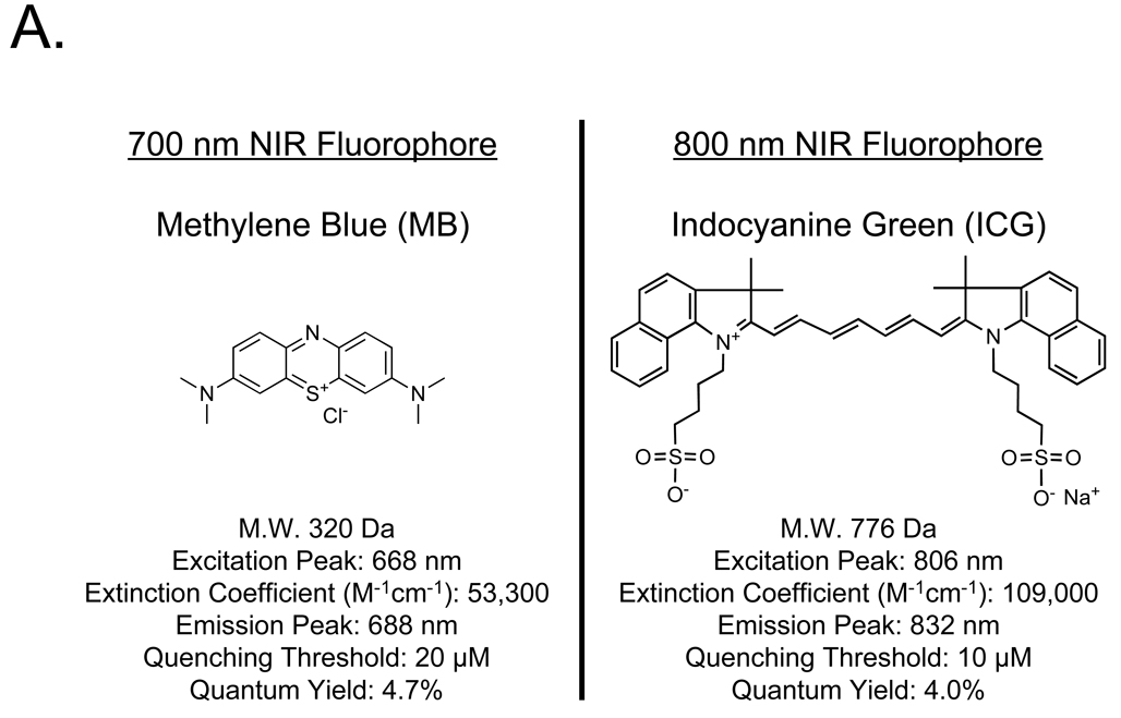
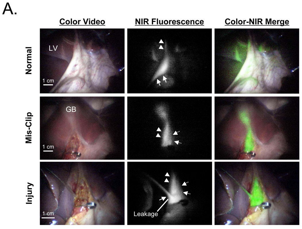
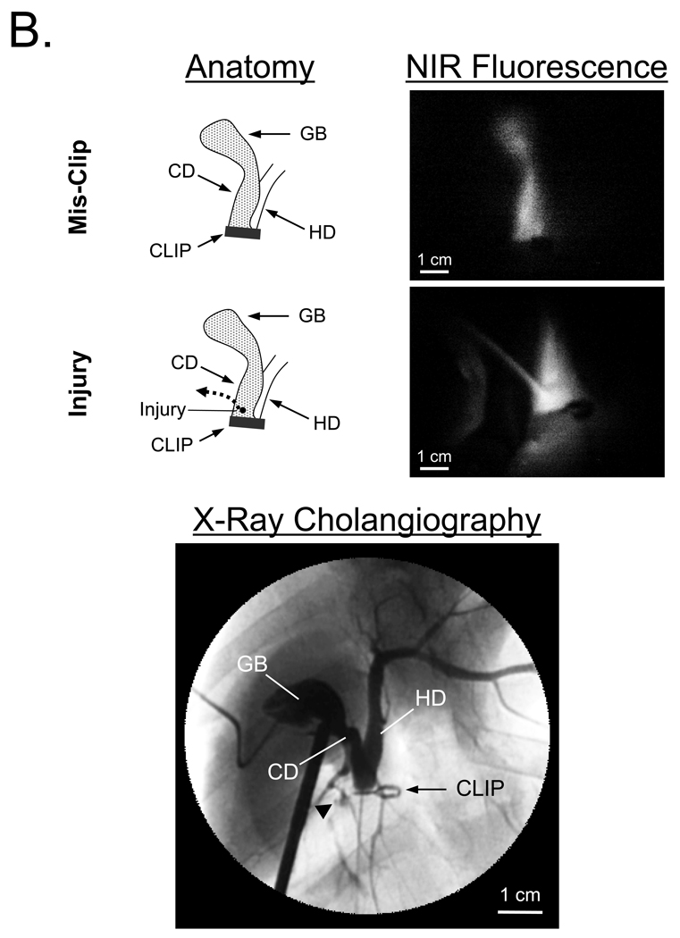
Background: Iatrogenic bile duct injuries are serious complications with patient morbidity. We hypothesized that the invisible near-infrared (NIR) fluorescence properties of methylene blue (MB) and indocyanine green (ICG) could be exploited for real-time, intraoperative imaging of the extrahepatic bile ducts during open and laparoscopic surgeries.

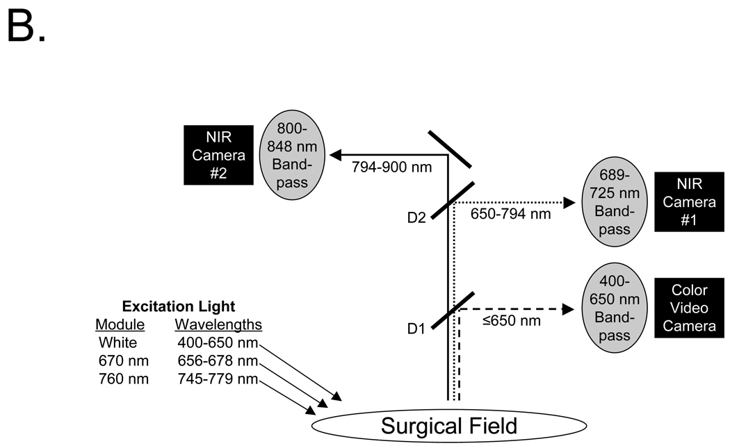
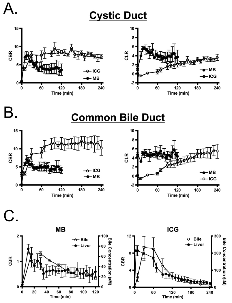
Methods: In all, 2.0 mg/kg of MB and 0.05 mg/kg of ICG were injected intravenously into 35-kg female Yorkshire pigs and the extrahepatic bile ducts were imaged over time using either the Fluorescence-Assisted Resection and Exploration (FLARE) image-guided surgery system (open surgery) or a custom NIR fluorescence laparoscopy system. Surgical anatomy was confirmed using x-ray cholangiography. The contrast-to-background ratio (CBR), contrast-to-liver ratio (CLR), and chemical concentrations in the cystic duct (CD) and common bile duct (CBD) were measured, and the performance of each agent was quantified.

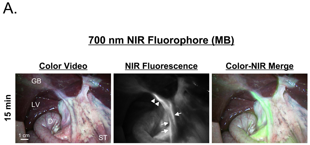
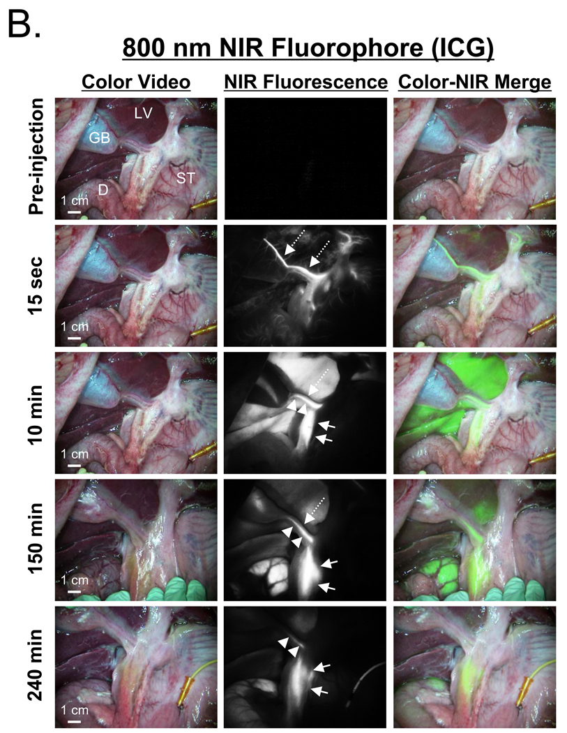
Results: Using NIR fluorescence of MB, the CD and CBD could be identified with good sensitivity (CBR and CLR > or =4), during both open and laparoscopic surgeries, from 10 to 120 min postinjection. Functional impairment of the ducts, including constriction and injury were immediately identifiable. Using NIR fluorescence of ICG, extrahepatic bile ducts did not become visible until 90 min postinjection because of strong residual liver retention; however, between 90 and 240 min, ICG provided exquisitely high sensitivity for both CD and CBD, with CBR > or =8 and CLR > or =4.

Conclusion: We demonstrate that 2 clinically available NIR fluorophores, MB fluorescing at 700 nm and ICG fluorescing at 800 nm, provide sensitive, prolonged identification of the extrahepatic bile ducts and assessment of their functional status.


Figures

Figure 1
Figure 1. NIR Fluorescent Contrast Agents and Imaging System Configuration
A. Chemical structures and optical properties of methylene blue (MB) and indocyanine green (ICG) in swine bile. B. The optical paths, dichroic mirrors (D1, D2), and filtration for the FLARE™ image-guided open surgery system used in this study. Filter wavelength ranges (nm) are provided for all excitation and emission filters. D1 = 680 nm center-wavelength dichroic mirror. D2 = 770 nm center-wavelength dichroic mirror.
Figure 2
Figure 2. NIR Fluorescence-Guided Intraoperative Identification of the Extrahepatic Bile Duct
Shown are the color video (left column), NIR fluorescence (middle column), and a pseudo-colored (lime green) merged image of the two (right column). Arrows, dashed arrows, and arrowheads indicate the common bile duct, cystic artery, and cystic duct, respectively. The cystic artery appears black at time points later than 15 sec post-injection. GB: gallbladder, LV: liver, ST: stomach, D: duodenum. A. 700 nm NIR fluorescence of 2.0 mg/kg MB at 15 min post-injection. Exposure time was 150 msec for the NIR fluorescence image. B. 800 nm NIR fluorophore 0.05 mg/kg ICG at the post-injection times indicated. Exposure time was 30 msec for all NIR fluorescence images.
Figure 3
Figure 3. Quantitative Assessment of Extrahepatic Bile Duct Imaging using NIR Fluorescence
A. Direct comparison of CBR (mean ± SEM; left) and CLR (mean ± SEM; right) between MB (closed circles) and ICG (open circles) in the cystic duct. CBR and CLR were obtained from images taken with an exposure time of 150 msec for MB and 30 msec for ICG. B. Direct comparison of CBR (mean ± SEM; left) and CLR (mean ± SEM; right) between MB (closed circles) and ICG (open circles) in the common bile duct, as described in Figure 3A. C. Correlation between imaging CBR (mean ± SEM) and actual NIR fluorophore concentration (mean ± SEM) in the liver (closed circles) and bile (open circles) for MB (left) and ICG (right). All image pixel intensities were within the linear range of the cameras.
Figure 4
Figure 4. Intraoperative NIR Fluorescence-Guided Extrahepatic Bile Duct Imaging during Laparoscopy
A. Laparoscopic NIR fluorescence-guided extrahepatic bile duct imaging. Images include color video (left column), NIR fluorescence (middle column), and a pseudo-colored (lime green) merged image of the two (right column). Top row images are from 10 min post-injection of 2 mg/kg MB, with the gallbladder retracted upward, and out of, the field of view. Middle row images present the common bile duct mis-clipped at slightly below the origin of the cystic duct. Bottom row images show an injury to the common bile duct and leakage of MB solution from the injured hole. All NIR fluorescence images were taken with an exposure time 500 msec. Arrow, dashed arrow, and arrowhead indicate the common bile duct, common hepatic duct, and cystic duct, respectively. LV: liver, GB: gallbladder. B. Verification of the surgical anatomy seen using NIR fluorescence laparoscopy by x-ray cholangiography. Schematic and NIR fluorescence images after mis-application of a surgical clip and after induction of CBD injury (top). At the bottom is shown the x-ray cholangiogram of the same animal after intra-GB injection of 5 ml of Renografin®-60. Black arrowhead indicates leakage of iodine contrast. GB: gallbladder, CBD: common bile duct, CD: cystic duct, HD: hepatic duct.

Similar articles

Cited by

References

    1. Advance Data No. 385, Table 8. Number of all-listed procedures for discharges from short-stay hospitals by procedure category and age: United States, 2005. U.S. Department of Health and Human Services, Centers for Disease Control and Prevention, National Center for Health Statistics; 2007.
    1. Flum DR, Dellinger EP, Cheadle A, Chan L, Koepsell T. Intraoperative cholangiography and risk of common bile duct injury during cholecystectomy. Jama. 2003;289:1639–1644. - PubMed
    1. Nuzzo G, Giuliante F, Giovannini I, Ardito F, D'Acapito F, Vellone M, et al. Bile duct injury during laparoscopic cholecystectomy: results of an Italian national survey on 56 591 cholecystectomies. Arch Surg. 2005;140:986–992. - PubMed
    1. Savassi-Rocha PR, Almeida SR, Sanches MD, Andrade MA, Frerreira JT, Diniz MT, et al. Iatrogenic bile duct injuries. Surg Endosc. 2003;17:1356–1361. - PubMed
    1. Buanes T, Mjaland O, Waage A, Langeggen H, Holmboe J. A population-based survey of biliary surgery in Norway. Relationship between patient volume and quality of surgical treatment. Surg Endosc. 1998;12:852–855. - PubMed

Publication types