. 2010 Jul 1;87(1):40-9.
doi: 10.1093/cvr/cvq027. Epub 2010 Jan 29.

Transplantation of cardiac progenitor cell sheet onto infarcted heart promotes cardiogenesis and improves function

Affiliations

Transplantation of cardiac progenitor cell sheet onto infarcted heart promotes cardiogenesis and improves function

Liudmila Zakharova et al. Cardiovasc Res. .

Abstract

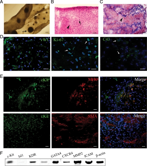
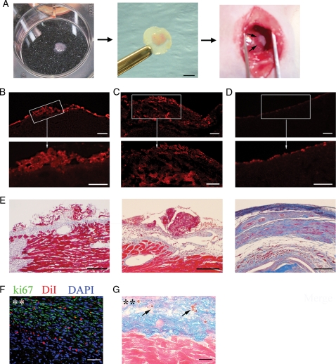
Aims: Cell-based therapy for myocardial infarction (MI) holds great promise; however, the ideal cell type and delivery system have not been established. Obstacles in the field are the massive cell death after direct injection and the small percentage of surviving cells differentiating into cardiomyocytes. To overcome these challenges we designed a novel study to deliver cardiac progenitor cells as a cell sheet.

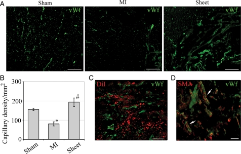
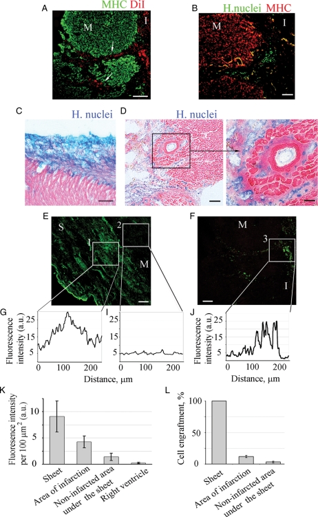
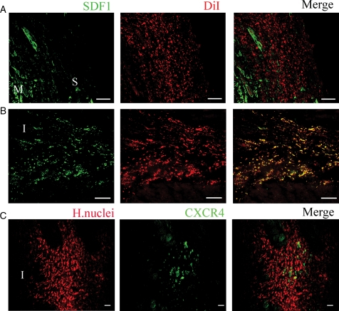
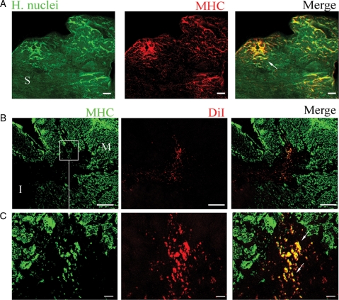
Methods and results: Cell sheets composed of rat or human cardiac progenitor cells (cardiospheres), and cardiac stromal cells were transplanted onto the infarcted myocardium after coronary artery ligation in rats. Three weeks later, transplanted cells survived, proliferated, and differentiated into cardiomyocytes (14.6 +/- 4.7%). Cell sheet transplantation suppressed cardiac wall thinning and increased capillary density (194 +/- 20 vs. 97 +/- 24 per mm(2), P < 0.05) compared with the untreated MI. Cell migration from the sheet was observed along the necrotic trails within the infarcted area. The migrated cells were located in the vicinity of stromal-derived factor (SDF-1) released from the injured myocardium, and about 20% of these cells expressed CXCR4, suggesting that the SDF-1/CXCR4 axis plays, at least, a role in cell migration. Transplantation of cell sheets resulted in a preservation of cardiac contractile function after MI, as was shown by a greater ejection fraction and lower left ventricular end diastolic pressure compared with untreated MI.

Conclusion: The scaffold-free cardiosphere-derived cell sheet approach seeks to efficiently deliver cells and increase cell survival. These transplanted cells effectively rescue myocardium function after infarction by promoting not only neovascularization but also inducing a significant level of cardiomyogenesis.


Figures

Figure 1
Figure 1
Cell sheet characteristics. (A) Fully formed cell sheet. Arrow indicates integrated cardiosphere. (B) H&E staining; pink colour (arrowhead) indicates cytosol and blue (arrows) indicates nuclear stain. Note that there is no necrotic core within the cell sheet. (C) Masson's Trichrome staining of sheet section. Arrowhead indicates collagen deposition within the sheet. (D and E) Sheet sections were labelled with antibodies against following markers: (D) vWf (green), Ki-67 (green), C43 (green); (E) c-Kit (green), MHC (red), SMA (red) as indicated on top of each panel. Nuclei were labelled with blue fluorescence of 4′,6-diamidino-2-phenylindole (DAPI). (F) Gene expression analysis of the cell sheet. Scale bars, 200 μm (A) or 50 μm (B–E).
Figure 2
Figure 2
Transplantation and growth of cell sheet after transplantation. (A) Sheet transplantation onto infarcted heart. Detached cell sheet on six-well plate (left); cell sheet folded on filter (middle); and transplanted onto left ventricle (right). Scale bar 2 mm. DiI-labelled cell sheets grafted above MI area at day 3 (B) and day 21 (C) after transplantation. (D) LV section of untreated MI rat at day 21 showing no significant red fluorescence background. Bottom row (B–D) demonstrates the enlargement of box-selected area of corresponding top panels. (E) Similar sections stained with Masson's Trichrome. Section of rat (F) or human (G) sheet treated rat at day 21 after MI. (F) Section was stained with antibody against Ki-67 (green). Cell sheet was pre-labelled with DiI (red). Nuclei stained with blue fluorescence of DAPI. (G) Section was double stained with human nuclei (blue) and caspase 3 (brown, arrows) antibodies and counterstained with eosin. Asterisks (**) indicate cell sheet area. Scale bars 200 μm (B–D, top row), 100 μm (B–D, bottom row, and E) or 50 μm (F, G).
Figure 3
Figure 3
Neovascularization of infarcted wall. (A) Frozen tissue sections stained with vWf antibody (green). LV section of control (sham), infarcted (MI), and MI treated with cell sheet (sheet) rats. Scale bar, 100 μm. (B) Capillary density decreased in the MI compared with sham (*P < 0.05) and improved after cell sheet treatment (#P < 0.05). (C) Neovascularization within cell sheet area was recognized by co-localization of DiI- (red) and vWf (green) staining. Scale bar 100 μm. (D) Mature blood vessels (arrows) were identified by co-localization of SMA (red) and vWf (green) staining. Scale bar 50 μm.
Figure 4
Figure 4
Engraftment quantification of cells migrated from the sheet into the infarcted area of MI. Animals were treated with rat (A) or human (BF) sheets. Cardiomyocytes were labelled with MHC antibody (A, green or B, red). Rat sheet-originated cells were identified with DiI-labelling, red (A). Arrows indicate the track of migrating cells. Human sheet-originated cells were identified by immunostaining with human nuclei antibody followed by secondary antibodies conjugated with either Alexa 488 (B, E and F, green) or AP (C, D, blue). No migration was detected when the cell sheet was transplanted onto non-infarcted myocardium (C). Heart sections were counterstained with eosin, pink (C–D). Higher magnification of area selected in the box is presented (D, right). Immunofluorescence of sheet (green) grafted to the myocardium surface (E) or cells migrated to the infarction area (F). Fluorescence profiles across the cell sheet itself (G, box 1), area underlying cell sheet (I, box 2) and infarction area with migrated cells (F, box 3). Mean fluorescence intensity of the grafted human (K) cells was determined by outlining the region of interest (ROI) and subtracting the background fluorescence for the same region. Fluorescence intensity was normalized to the area of ROI (n = 6). (L) Percent engraftment was defined as number of lamin-positive cells divided by total number of cells per ROI. ‘M’, myocardium,’S’ sheet, ‘I’ infarction. Scale bars 100 μm (A–C, D, left, E and F), or 50 μm (D, right).
Figure 5
Figure 5
Migration of sheet-originated cells into the infarcted area. Confocal images of MI animals treated with sheets from rats (A and B) or human (C). SDF1 (green) was detected at border zone of the infarct at day 3 (A) and day 21 (B). Rat sheet-originated cells were identified with DiI-labelling (red). Note co-localization of DiI-positive sheet-originated cells with SDF1 at 21 days after MI (B). Human cells were identified by immunostaining with human nuclei antibody, red, (C). Note human cells that migrated to the area of infarct express CXCR4 (green) (C). Scale bar, 200 μm (A, B) or 50 μm (C). ‘M’, myocardium, ‘S’ sheet, ‘I’ infarct.
Figure 6
Figure 6
Cardiac regeneration. Sections of MI animals treated with human (A) or rat (B, C) sheets. Human sheet was identified by immunostaining with human nuclei antibody (green). Section was double-stained with MHC (red) antibody. Newly formed cardiomyocytes was identified by co-localization of human nuclei and MHC (yellow, arrow). (B) Rat sheet-originated cells were identified by DiI labelling (red). Section was double-stained with MHC (green) antibody. Newly formed cardiomyocytes were detected by co-localization of DiI with MHC (yellow, arrows). (C) Higher magnification of area selected in the boxes (B). Scale bars 200 μm (B), or 20 μm (A, C). ‘M’, myocardium, ‘S’ sheet, ‘I’ infarct.

Comment in

References

    1. Fuchs JR, Nasseri BA, Vacanti JP, Fauza DO. Postnatal myocardial augmentation with skeletal myoblast-based fetal tissue engineering. Surgery. 2006;140:100–107. - PubMed
    1. Orlic D, Kajstura J, Chimenti S, Bodine DM, Leri A, Anversa P. Bone marrow stem cells regenerate infarcted myocardium. Pediatr Transplant. 2003;7(Suppl. 3):86–88. - PubMed
    1. Kawamoto A, Tkebuchava T, Yamaguchi J, Nishimura H, Yoon YS, Milliken C, et al. Intramyocardial transplantation of autologous endothelial progenitor cells for therapeutic neovascularization of myocardial ischemia. Circulation. 2003;107:461–468. - PubMed
    1. Iwasaki H, Kawamoto A, Ishikawa M, Oyamada A, Nakamori S, Nishimura H, et al. Dose-dependent contribution of CD34-positive cell transplantation to concurrent vasculogenesis and cardiomyogenesis for functional regenerative recovery after myocardial infarction. Circulation. 2006;113:1311–1325. - PubMed
    1. Beltrami AP, Barlucchi L, Torella D, Baker M, Limana F, Chimenti S, et al. Adult cardiac stem cells are multipotent and support myocardial regeneration. Cell. 2003;114:763–776. - PubMed

Publication types

MeSH terms

Substances