. 2010 Dec;43(6):731-9.
doi: 10.1165/rcmb.2009-0328OC. Epub 2010 Jan 29.

Temporal relationship between primary and motile ciliogenesis in airway epithelial cells

Affiliations

Temporal relationship between primary and motile ciliogenesis in airway epithelial cells

Raksha Jain et al. Am J Respir Cell Mol Biol. 2010 Dec.

Abstract

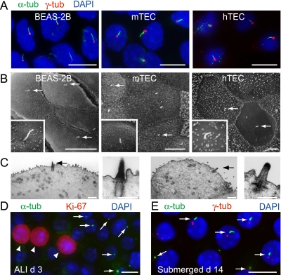
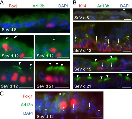
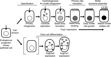
Cilia are traditionally classified as motile or primary. Motile cilia are restricted to specific populations of well-differentiated epithelial cells, including those in the airway, brain ventricles, and oviducts. Primary cilia are nonmotile, solitary structures that are present in many cell types, and often have sensory functions such as in the retina and renal tubules. Primary cilia were also implicated in the regulation of fundamental processes in development. Rare depictions of primary cilia in embryonic airways led us to hypothesize that primary cilia in airway cells are temporally related to motile ciliogenesis. We identified primary cilia in undifferentiated, cultured airway epithelial cells from mice and humans and in developing lungs. The solitary cilia in the airways express proteins considered unique to primary cilia, including polycystin-1 and polycystin-2. A temporal analysis of airway epithelial cell differentiation showed that cells with primary cilia acquire markers of motile ciliogenesis, suggesting that motile ciliated cells originate from primary ciliated cells. Whereas motile ciliogenesis requires Foxj1, primary ciliogenesis does not, and the expression of Foxj1 was associated with a loss of primary cilia, just before the appearance of motile cilia. Primary cilia were not found in well-differentiated airway epithelial cells. However, after injury, they appear in the luminal layer of epithelium and in basal cells. The transient nature of primary cilia, together with the temporal and spatial patterns of expression in the development and repair of airway epithelium, suggests a critical role of primary cilia in determining outcomes during airway epithelial cell differentiation.


Figures

Figure 1.
Figure 1.
Primary cilia are present on primary culture airway epithelial cells. (A) Human airway epithelial cell line BEAS-2B and tracheal epithelial cells from mice and humans (mTEC and hTEC), cultured using air–liquid interface (ALI) conditions, were immunostained with antibodies for cilia protein acetylated α-tubulin (α-tub, green) and basal body protein γ-tubulin (γ-tub, red). Nuclei were stained using 4′,6 diamidino-2-phenylindole (DAPI) (blue). Overlaid photomicrographs of BEAS-2B and preparations on ALI Day 2 (mTEC) and Day 1 (hTEC) demonstrate primary cilia. (B) Scanning electron microscopy (EM) of preparations (as in A) show solitary primary cilia. Bars = 5 μm. (C) Transmission EM of mTEC preparations on ALI Day 3 show solitary primary cilia on the cell surface (arrows). Bars = 1 μm at low magnification, and 0.5 μm at high magnification. (D) mTEC preparations on ALI Day 3, immunostained for α-tub (arrows) and cell-cycle protein Ki-67 (arrowheads), show primary cilia on cells that lack Ki-67 expression. (E) Primary cilia (arrows) are abundant in mTEC preparations cultured for 14 days using submerged rather than ALI conditions, immunostained as in A. Bars in A, D, and E = 10 μm.
Figure 2.
Figure 2.
Polycystin-1 and polycystin-2 are expressed in primary and motile cilia of airway epithelial cells. (A) Preparations of primary-culture tracheal epithelial cells were immunostained for cilia marker acetylated α-tubulin (α-tub) and polycystin-1 or polycystin-2 (PC-1 or PC-2) on ALI Day 10 (hTEC) or Day 1 (mTEC). Overlaid photomicrographs show colocalization of polycystins with α-tub in primary cilia. PC-1 and PC-2 were not detected in cilia incubated with normal rabbit IgG. (B) Well-differentiated mTEC preparations immunostained for PC-2 and α-tub demonstrate co-expression in motile cilia, shown en face (top) and in isolated cells (bottom). Bars in A and B = 10 μm. (C) Protein blot analysis of primary culture tracheal cell preparations, harvested on indicated day of ALI culture, were analyzed for expression of PC-1 and PC-2, α-tub, cilia protein Arl13b, and motile ciliated cell transcription factor Foxj1. Samples from mouse kidney (mkidney) and MDCK cells are indicated. Membranes were reprobed to detect the indicated protein. PC-1 was detected on a separate membrane, to resolve a protein of approximately 500 kD.
Figure 3.
Figure 3.
Primary cilia appear in airway epithelial cells before motile ciliogenesis in vitro and in developing lungs. (A) Preparations of mTEC were analyzed on indicated day of ALI culture by immunostaining, using antibodies for acetylated α-tubulin (α-tub, green) and γ-tubulin (γ-tub, red). Cells with primary cilia on ALI Day 1 decreased in abundance as motile cilia appeared on ALI Day 4, and as cells became well-differentiated on ALI Day 14. (B) Mouse lung sections from embryonic day (E) 12.5 were immunostained with antibodies to α-tub (green) and Arl13b (red), and show primary cilia. (C) Transition from primary to motile cilia during development. Lungs on E14.5 and E15.5 were immunostained for primary and motile cilia protein Arl13b (green) and motile cilia protein β-tubulin–IV (β-tub-IV, red). Insets show detail. Primary cilia (arrows) and motile cilia (arrowheads) are indicated. All bars = 10 μm.
Figure 4.
Figure 4.
Primary ciliogenesis is Foxj1-independent. (A) mTEC derived from wild-type (WT) and Foxj1−/− mice, cultured under ALI conditions, were immunostained for acetylated α-tubulin (α-tub, green) and γ-tubulin (γ-tub, red). On ALI Day 2, primary cilia are similar in mTECs from each strain. On ALI Day 14, Foxj1−/− cells show clusters of basal bodies (red) that fail to dock at the apical membrane (arrows, lower right) and primary cilia, unlike the motile cilia in WT mTECs. (B) Primary cilia in Foxj1-expressing cells during lung development. Lung sections from the indicated embryonic day (E) were immunostained for Arl13b (green) and Foxj1 (red). On E14.5, primary cilia are present before Foxj1 expression. At the onset of Foxj1 expression, on E15.5, primary cilia are present in some Foxj1-expressing cells, and absent in others. On E19.5, motile-type cilia are present on nearly all Foxj1 expressing cells (insets show detail). Representative airways with primary (arrows) and motile (arrowheads) cilia are shown. All bars = 10 μm.
Figure 5.
Figure 5.
Motile ciliated cells originate from primary ciliated cells. (A) mTEC preparations were immunostained for acetylated α-tubulin on the indicated ALI day, and ciliated cell types were quantified. Shown are the means ± SDs of cilia types in three random fields of supported membranes from at least three independent preparations (*P < 0.05, primary versus motile ciliated cells). (B) Primary cilia are lost during early motile ciliogenesis in mTECs (ALI Days 1–4). Primary cilia (Arl13b, red) are evident in cells before the expression of Foxj1 (green) (Day 1). Primary cilia are evident in Foxj1-expressing cells at time of onset of Foxj1 expression (Day 2). Loss of primary cilia occurs in cells that express Foxj1 as motile ciliogenesis proceeds (Day 3), and motile cilia develop (Day 4). (C) Quantification of cilia type in cells that express Foxj1 in B. Shown are the means ± SDs of Foxj1-expressing cells in one inset each day, from each of four independent preparations. Foxj1-expressing cells were evaluated for the totals: Day 2, n = 795; Day 3, n = 1,184; and Day 4, n = 1,413. (D) Primary cilia (α-tub, green) in mTECs undergoing early motile ciliogenesis marked by clusters of centrioles (basal bodies, γ-tub, red) within the cytoplasm (ALI Day 2). Primary cilia are absent after the docking of basal bodies and before motile cilia formation (ALI Day 3). (E) In the developing lung, primary cilia (α-tub, green) are absent in E16.5 cells that express early Clara cell marker Scgb3a2 (red; adjacent cell with cilium is outlined) and in adult cells that express Scgb1a1 (red, Clara cell secretory protein). (F) Scanning EM of adult mouse airways shows an absence of primary cilia in nonciliated cells, with the typical domed appearance of Clara cells. All bars = 10 μm.
Figure 6.
Figure 6.
Primary cilia are transiently present in adult mouse airway during repair after respiratory virus injury. Lung sections from mice were administered Sendai virus (SeV), harvested on the indicated day after infection. (A) Cilia type relative to Foxj1 expression was determined by immunostaining for Arl13b (green) and Foxj1 (red). On Day 8, SeV-infected mice lack cilia and Foxj1 expression. On Day 12, Foxj1-expressing cells have primary cilia (arrows) or no cilia. On Day 21, Foxj1-expressing cells have motile cilia (arrowheads). (B) Primary cilia in basal cells were determined by immunostaining for Arl13b (green) and basal cell maker cytokeratin 14 (K14, red). On Day 8, K14-expressing cells are not present. On Days 12 and 15, primary cilia are found in K14-expressing cells. By Day 21, motile cilia and fewer K14-expressing cells are present in airways. (C) Basal cells with primary cilia (arrows) are independent of Foxj1-expressing cells (immunostained as in A). Representative airways are shown. Bars = 10 μm.
Figure 7.
Figure 7.
Proposed model for relationship of primary cilia to motile ciliogenesis during airway epithelial cell differentiation. A progenitor airway epithelial cell with primary cilia generates daughter cells that enter motile cilia and secretory cell lineage pathways. Within the appropriate environment, the presence of a primary cilium marks cells that generate multiple centrioles (nascent basal bodies) and then express Foxj1, indicating commitment to a motile ciliated cell. As motile ciliogenesis proceeds to a high Foxj1 state, primary cilia are lost. In the Clara cell lineage, an undefined cell (in brackets) differentiates cells identified by markers of early commitment (Scgb3a2) and maturation (Scgb1a1, CCSP) that lack a primary cilium.

Similar articles

Cited by

References

    1. Badano JL, Mitsuma N, Beales PL, Katsanis N. The ciliopathies: an emerging class of human genetic disorders. Annu Rev Genomics Hum Genet 2006;7:125–148. - PubMed
    1. Fliegauf M, Benzing T, Omran H. When cilia go bad: cilia defects and ciliopathies. Nat Rev Mol Cell Biol 2007;8:880–893. - PubMed
    1. Gerdes JM, Davis EE, Katsanis N. The vertebrate primary cilium in development, homeostasis, and disease. Cell 2009;137:32–45. - PMC - PubMed
    1. Pazour GJ, Witman GB. The vertebrate primary cilium is a sensory organelle. Curr Opin Cell Biol 2003;15:105–110. - PubMed
    1. Davenport JR, Yoder BK. An incredible decade for the primary cilium: a look at a once-forgotten organelle. Am J Physiol Renal Physiol 2005;289:F1159–F1169. - PubMed

Publication types

MeSH terms

Substances