. 2011 Jan;44(1):34-9.
doi: 10.1165/rcmb.2009-0005OC. Epub 2010 Jan 29.

Micromechanics of alveolar edema

Affiliations

Micromechanics of alveolar edema

Carrie E Perlman et al. Am J Respir Cell Mol Biol. 2011 Jan.

Abstract

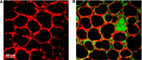
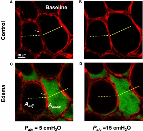
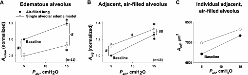
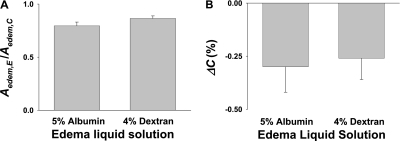
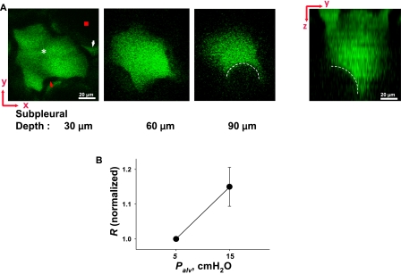
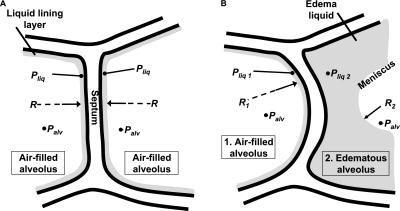
The decrease of lung compliance in pulmonary edema underlies ventilator-induced lung injury. However, the cause of the decrease in compliance is unknown. We tested the hypothesis that in pulmonary edema, the mechanical effects of liquid-filled alveoli increase tissue stress in adjacent air-filled alveoli. By micropuncture of isolated, perfused rat lungs, we established a single-alveolus model of pulmonary edema that we imaged using confocal microscopy. In this model, we viewed a liquid-filled alveolus together with its air-filled neighbor at different transpulmonary pressures, both before and after liquid-filling. Instilling liquid in an alveolus caused alveolar shrinkage. As a result, the interalveolar septum was stretched, causing the neighboring air-filled alveolus to bulge. Thus, the air-filled alveolus was overexpanded by virtue of its adjacency to a liquid-filled alveolus. Confocal microscopy at different depths of the liquid-filled alveolus revealed a meniscus. Lung inflation to near-total lung capacity (TLC) demonstrated decreased compliance of the air-filled but not liquid-filled alveolus. However, at near TLC, the air-filled alveolus was larger than it was in the pre-edematous control tissue. In pulmonary edema, liquid-filled alveoli induce mechanical stress on air-filled alveoli, reducing the compliance of air-filled alveoli, and hence overall lung compliance. Because of increased mechanical stress, air-filled alveoli may be susceptible to overdistension injury during mechanical ventilation of the edematous lung.


Figures

Figure 1.
Figure 1.
Low-power images of alveolar field. (A) Air-filled lung with calcein red–orange loaded into alveolar epithelial cells. Epithelium fluoresces red; alveolar liquid lining layer and air appear black. (B) Single alveolar edema model. Calcein red–orange labels the alveolar epithelium. Albumin solution labeled with green fluorescent 2′,7′-bis-(2-carboxyethyl)-5-(and-6)-carboxyfluorescein (BCECF) fills a single alveolus. All other alveoli are air-filled, with BCECF labeling the liquid lining layer. Images were obtained with a ×10 air objective.
Figure 2.
Figure 2.
Single alveolar edema model. Optical sections (2 μm thick) of two alveoli at a depth of 20 μm below the pleural surface. Alveolar epithelium is labeled with calcein red–orange at transpulmonary pressures (Palv) of 5 (A, C), and 15 (B, D) cm H2O. Alveoli are air-filled (Control) or liquid-filled with albumin solution (Edema), as indicated. White arrows indicate septum separating the two alveoli. Yellow scale marks indicate alveolar diameters at Palv of 5 cm H2O in control condition. Aedem, cross-sectional area of right alveolus that becomes liquid-filled. Aadj, cross-sectional area of adjacent left alveolus that remains air-filled.
Figure 3.
Figure 3.
Pressure-induced alveolar expansion. Cross-sectional area data for alveolar air pressures (Palv) of 5 and 15 cm H2O. Lines connecting data points at different Palv are inflation curves, with slopes indicating degree of pressure-induced alveolar expansion. Solid circles, before induction of single-alveolar edema model. Open circles, after induction of edema model. (A) Data for cross-sectional area (Aedem) of the alveolus that became liquid-filled. All four data points were obtained in the same alveoli (n = 11, from nine different lungs). (B) Data for cross-sectional area (Aadj) of the alveolus adjacent to the one that became liquid-filled. All four data points were obtained in the same alveoli (n = 13, from eight different lungs). (C) Aadj data for a representative individual alveolus. Combining all data in the air-filled lung before liquid-filling an alveolus: inflation increased alveolar cross-sectional area A from 7.0 ± 0.6 × 103 μm2 by 23% ± 2%, and alveolar perimeter length L from 3.2 ± 0.1 × 102 μm by 8% ± 1% (P < 0.01, n = 24). *Area greater at Palv of 15 than 5 cm H2O (P < 0.01), for either control air-filled state or edema model. #Area different in edema model than air-filled state, at constant Palv (P < 0.01). ##Area different in edema model than in air-filled state, at constant Palv (P < 0.02). Slope less in edema model than in air-filled state (P < 0.01), indicating decreased compliance.
Figure 4.
Figure 4.
Effect of albumin on alveolar mechanics. (A) Effect on liquid-filling induced decrease in alveolar cross-sectional area. Data comprise ratios of area values in edematous state after liquid-filling (Aedem,E) to those in control state before liquid-filling (Aedem,C). (B) Liquid-filling induced changes in compliance (ΔC(%)) of alveolus that became edematous. Albumin solution: n = 11 alveoli from nine lungs. Dextran solution: n = 9 alveoli from eight lungs.
Figure 5.
Figure 5.
Meniscus observed in edematous alveolus. (A) Left, 2-μm-thick optical sections (x–y plane) of an edematous alveolus at indicated depths below the pleural surface, at Palv of 15 cm H2O. Edema liquid (white asterisk) and liquid lining layer of neighboring alveolus (white arrowhead) are fluorescent. Alveolar wall (red arrowhead) and air-filled lumen of neighboring alveolus (red square) appear black. Starting at ∼ 85 μm below the pleural surface, a black crescent (dashed white line) bulges into the edema liquid. Right, y–z plane reconstructed from multiple sections. Dashed white line indicates meniscus. (B) Inflation increases meniscus radius (R) (P < 0.05). Data are paired, and normalized by baseline value of 32 ± 2 μm (n = 9).
Figure 6.
Figure 6.
Model of trans-septal forces. (A) Model of septum between two alveoli in normal, air-filled lung. In each alveolus, airway pressure is indicated as Palv. A thin liquid layer with liquid pressure (Pliq) lines each alveolus. With all forces balanced across the septum, the septum is planar. Radius (R) of air–liquid interface in each alveolus is thus infinite. (B) Model of septum between an air-filled alveolus (1) and an edematous alveolus (2). In each alveolus, airway pressure is indicated as Palv. Because of the presence of a meniscus, however, R2 in the edematous alveolus is less than R1 in the air-filled alveolus. According to the Laplace relationship, a greater pressure drop across the air–liquid interface occurs in the edematous than in the air-filled alveolus. Thus, liquid phase pressure in the edematous alveolus (Pliq2) is lower than Pliq1 in the air-filled alveolus. The difference in liquid phase pressures acting across the septum displaces the septum toward the edematous alveolus. Thus, liquid-filling diminishes the edematous alveolus, and expands the adjacent air-filled alveolus.

References

    1. Staub NC, Nagano H, Pearce ML. Pulmonary edema in dogs, especially the sequence of fluid accumulation in lungs. J Appl Physiol 1967;22:227–240. - PubMed
    1. Bachofen H, Schurch S, Michel RP, Weibel ER. Experimental hydrostatic pulmonary edema in rabbit lungs. Morphology. Am Rev Respir Dis 1993;147:989–996. - PubMed
    1. Perlman CE, Bhattacharya J. Alveolar expansion imaged by optical sectioning microscopy. J Appl Physiol 2007;103:1037–1044. - PubMed
    1. Weibel ER, Gomez DM. Architecture of the human lung. Science 1962;137:577–585. - PubMed
    1. Von Neergaard K. Neue Auffassungen über einen Grundbegriff der Atemmechanik. die Retraktionskraft der Lunge, abhangig von der Oberflachenspannung in den Alveolen. Z Gesamte Exp Med 1929;66:373–394.

Publication types

MeSH terms