. 2010 Apr 2;285(14):10454-63.
doi: 10.1074/jbc.M109.093427. Epub 2010 Jan 29.

A conserved threonine residue in the juxtamembrane domain of the XA21 pattern recognition receptor is critical for kinase autophosphorylation and XA21-mediated immunity

Affiliations

A conserved threonine residue in the juxtamembrane domain of the XA21 pattern recognition receptor is critical for kinase autophosphorylation and XA21-mediated immunity

Xuewei Chen et al. J Biol Chem. .

Abstract

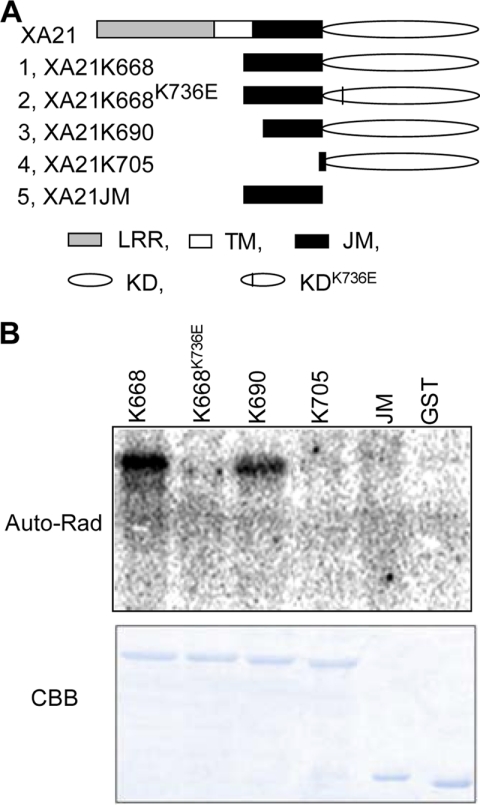
Despite the key role that pattern recognition receptors (PRRs) play in regulating immunity in plants and animals, the mechanism of activation of the associated non-arginine-aspartate (non-RD) kinases is unknown. The rice PRR XA21 recognizes the pathogen-associated molecular pattern, Ax21 (activator of XA21-mediated immunity). Here we show that the XA21 juxtamembrane (JM) domain is required for kinase autophosphorylation. Threonine 705 in the XA21 JM domain is essential for XA21 autophosphorylation in vitro and XA21-mediated innate immunity in vivo. The replacement of Thr(705) by an alanine or glutamic acid abolishes XA21 autophosphorylation and eliminates interactions between XA21 and four XA21-binding proteins in yeast and rice. Although threonine residues analogous to Thr(705) of XA21 are present in the JM domains of most RD and non-RD plant receptor-like kinases, this residue is not required for autophosphorylation of the Arabidopsis RD RLK BRI1 (brassinosteroid insensitive 1). The threonine 705 of XA21 is conserved only in the JM domains of plant RLKs but not in those of fly, human, or mouse suggesting distinct regulatory mechanisms. These results contribute to growing knowledge regarding the mechanism by which non-RD RLKs function in plant.


Figures

FIGURE 1.
FIGURE 1.
Kinase autophosphorylation assay for the XA21 variants. A, the structures of XA21 variants. B, the autophosphorylation assay for the E. coli-expressed, GST-fused XA21 variant proteins. The upper panel shows the autoradiograph (auto-rad) and the lower panel is a photograph of the gel stained with Coomassie Brilliant Blue (CBB). K668, K668K736E, K690, K705, and JM denote GST-XA21K668, GST-XA21K668K736E, GST-XA21K690, GST-XA21K705, and GST-XA21K668JM, respectively, and GST denotes GST alone.
FIGURE 2.
FIGURE 2.
Kinase autophosphorylation assay for the GST-XA21K668 variants. The autophosphorylation assay was performed for the E. coli-expressed, GST-fused XA21K668 variant proteins. The upper panel shows the autoradiograph (autorad) and the lower panel is a photograph of the gel stained with Coomassie Brilliant Blue (CBB). K668, K668K736E, K668S697A, K668S697D, K668T705A, K668T705E, K668T680A, K668S686A/T688A/S689A, and K668S699A, denote their XA21 GST fusion proteins, respectively.
FIGURE 3.
FIGURE 3.
Kinase autophosphorylation analyses of XA21K668 and its single amino acid mutants T705A and T705E. A, kinase autophosphorylation was determined for E. coli-expressed wild-type GST-XA21K668 (K668) and the GST-XA21K668T705A (T705A), and GST-XA21K668T705E (T705E) mutants. B, kinase autophosphorylation was determined for rice-expressed Myc-XA21 (XA21) and mutants Myc-XA21T705A (T705A) and Myc-XA21T705E (T705E). The upper panel in A show a Coomassie Brilliant Blue-stained gel and the upper panel in B shows a Western blot (WB) probed with an anti-Myc antibody to normalize protein loading. The middle panels show typical autoradiographs of the indicated proteins, and the bottom panels show quantification of kinase autophosphorylation. Error bars indicate the standard deviations from three independent experiments.
FIGURE 4.
FIGURE 4.
Test on the protein interactions of XA21K668 variants (XA21K668T705A and XA21K668T705E) with XBs in yeast. The cytoplasmic domain of XA21 (XA21K668), containing both the JM and kinase domains, and its variants XA21K668T705A and XA21K668T705E were used as baits in the yeast two-hybrid analysis. Upper panel, Western blot of yeast-expressed proteins. LexA-XA21K668 (K668), LexA-XA21K668T705A (T705A), and LexA-XA21K668T705E (T705E) proteins expressed in yeast were extracted and probed with an anti-LexA antibody. Lower panel, summary of protein interaction results. The XB proteins were fused to the activation domain. Blue color indicates the positive interaction.
FIGURE 5.
FIGURE 5.
Co-immunoprecipitation of XB15 and XB24 with Myc-XA21 and the mutant variants. A, co-immunoprecipitation of XB15. B, co-immunoprecipitation of XB24. Protein extracts from transgenic rice carrying wild-type Myc-XA21, Myc-XA21T705A, or Myc-XA21T705E were mixed with anti-Myc antibody coupled to agarose, and the associated proteins were precipitated. The precipitates were probed with anti-Myc, anti-XB15, or anti-XB24 antibody. Myc-XA21, Myc-XA21T705A, and Myc-XA21T705E proteins represent labeled XA21, T705A, and T705E, respectively.
FIGURE 6.
FIGURE 6.
Disease resistance evaluation of 6-week-old transgenic plants inoculated with PXO99AZ. The average disease length measured 14 days post-inoculation is shown in the upper panel. The middle panel shows the PCR-based genotyping for the presence of the Xa21 transgene on the transgenic plants and Myc-Xa21, and Kitaake control plants. Xa21a designates an Xa21-specific amplified fragment and NS indicates nonspecific amplification. The bottom panel shows RNA expression of transgenes detected in transgenic lines by reverse transcription-PCR. Xa21b indicates the amplification from Xa21 transcripts. The numbers in the middle and bottom panels represent independent transgenic lines. Xa21, T705A, and T705E represent transgenic plants carrying Myc-Xa21, Myc-Xa21T705A, and Myc-Xa21T705E, respectively. The numbers indicate the progeny lines from each T0 transgenic line used for the analyses. Kitaake is a control. Error bars represent the standard deviation.
FIGURE 7.
FIGURE 7.
Evaluation of transgenic plants carrying XA21, XA21T705A, or XA21T705E on disease resistance to the Xoo strain PXO99AZ. A, photograph of representative leaves taken 14 days after inoculation. B, lesion length development over time. Each data point represents the average and standard deviation of three leaves. C, bacterial growth curves. Each data point represents the average and standard deviation of three leaves. XA21, T705A, and T705E represent transgenic lines expressing wild-type Myc-XA21, Myc-XA21T705A, and Myc-XA21T705E, respectively. Kitaake is the rice recipient used in the transformation experiments.
FIGURE 8.
FIGURE 8.
Identification of Thr705 as an autophosphorylation site by mass spectrometry. GST-XA21K668 protein was incubated with or without the ATPase His-tagged XB24 (His-XB24). The purified GST-XA21K668 protein was subject to LC-MS/MS analysis. The top panel shows the spectrum of the peptide containing the Thr705 residue from the GST-XA21K668 protein subjected to kinase autophosphorylation in the presence of His-XB24. The spectrum was generated using Scaffold Viewer 2.0. T+80, indicated by the arrows, designates phosphorylated Thr705. The bottom panel summarizes the highest probability of phosphorylation of the peptide containing Thr705. GST-K668, GST-K668+ATP, and GST-K668+ATP+His-XB24 represent the different treatments of the GST-XA21K668 protein, namely GST-K668 only, GST-K668 subjected to kinase autophosphorylation, and GST-K668 subjected to kinase autophosphorylation in the presence of His-tagged XB24, respectively.
FIGURE 9.
FIGURE 9.
Threonine sites in the JM domain of selected plant receptor kinases analogous to Thr705 in XA21. A, the JM domains of rice RLKs XA21 (non-RD class), XA26 (non-RD class), and Pid2 (non-RD class), and Arabidopsis RLKs BRI1 (RD-class), FLS2 (non-RD class), and EFR (non-RD class) are listed. The JM sequences shown herein were identified using the SMART program. The boxed conserved threonine amino acid residues are two residues from the beginning of the core S/T kinase domain of all RLKs, except Pid2 (0 residues). B, analyses the equivalent sites to Thr705 of XA21 in all RLKs from the kinomes of rice and Arabidopsis. RD and non-RD represent RD and non-RD classes of RLKs, respectively. Os, Oryza sativa (rice); At, Arabidopsis thaliana.

Similar articles

Cited by

References

    1. Thiel K. W., Carpenter G. (2007) Proc. Natl. Acad. Sci. U.S.A. 104, 19238–19243 - PMC - PubMed
    1. Wang X., Li X., Meisenhelder J., Hunter T., Yoshida S., Asami T., Chory J. (2005) Dev. Cell 8, 855–865 - PubMed
    1. Agosti V., Karur V., Sathyanarayana P., Besmer P., Wojchowski D. M. (2008) Exp. Hematol. 37, 159–171 - PMC - PubMed
    1. Heiss E., Masson K., Sundberg C., Pedersen M., Sun J., Bengtsson S., Rönnstrand L. (2006) Blood 108, 1542–1550 - PubMed
    1. Aifa S., Frikha F., Miled N., Johansen K., Lundström I., Svensson S. P. (2006) Biochem. Biophys. Res. Commun. 347, 381–387 - PubMed

Publication types

MeSH terms