. 2010 Apr 2;285(14):10553-62.
doi: 10.1074/jbc.M109.065631. Epub 2010 Jan 29.

Activation of the nucleotide oligomerization domain signaling pathway by the non-bacterially derived xanthone drug 5'6-dimethylxanthenone-4-acetic acid (Vadimezan)

Affiliations

Activation of the nucleotide oligomerization domain signaling pathway by the non-bacterially derived xanthone drug 5'6-dimethylxanthenone-4-acetic acid (Vadimezan)

Guanjun Cheng et al. J Biol Chem. .

Abstract

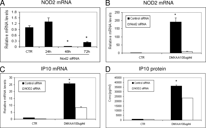
The cytosolic nucleotide-binding oligomerization domain 1 (NOD1)/CARD4 and NOD2/CARD15 proteins are members of NOD-like receptors recognizing specific motifs within peptidoglycans of both Gram-negative and Gram-positive bacteria. NOD1 and NOD2 signal via the downstream adaptor serine/threonine kinase RIP2/CARDIAK/RICK to initiate NF-kappaB activation and the release of inflammatory cytokines/chemokines. In this report, we show that 5,6-dimethylxanthenone-4-acetic acid (DMXAA), a cell-permeable, small molecule that has anti-tumor activity, can also activate NOD1 and NOD2. This was demonstrated: 1) by using human embryonic kidney epithelial (HEK) 293 cells transfected with a NF-kappaB reporter plasmid in combination with NOD1 or NOD2 expression plasmids; 2) by inhibiting DMXAA-induced chemokine (CXCL10) mRNA and protein production in the AB12 mesothelioma cell line using a pharmacological inhibitor of RICK kinase, SB20358; and 3) by using small interfering RNA to knock down NOD2 and lentiviral short hairpin RNA to knock down RICK. These findings expand the potential ligands for the NOD-like receptors, suggesting that other xanthone compounds may act similarly and could be developed as anti-tumor agents. This information also expands our knowledge on the mechanisms of action of the anti-tumor agent DMXAA (currently in clinical trials) and may be important for its biological activity.


Figures

FIGURE 1.
FIGURE 1.
Schematic representative of the chemical structures of MDP, iE-DAP, the flavone backbone, the parent flavanoid, FAA, DMXAA, and the xanthone parent backbone.
FIGURE 2.
FIGURE 2.
DMXAA stimulates NF-κB activity only in the presence of mNOD2, mNOD1. A, HEK293 cells were transiently transfected with an NF-κB firefly luciferase reporter plasmid. Some cells were co-transfected with a control plasmid (first three bars) or with an mNOD2 plasmid (last two bars). 18 h after transfection, cells were exposed to calcium phosphate alone or the NOD2 ligand, MDP (100 ng/ml), in conjunction with calcium phosphate to facilitate cell entry. After 24 h, NF-κB promoter activation was measured by firefly luciferase activity normalized to control cells. Each transfection was performed in triplicate. Data are shown as the mean ± S.E. for one of three representative experiments with similar results. A 5.2-fold increase in activity was seen in the mNOD2 + MDP + calcium group (*, p < 0.01, versus all other groups). B, HEK293 cells were transiently co-transfected NF-κB firefly luciferase reporter plasmid and with control vector (first two bars) or mNOD2 plasmid (last two bars) as above. 18 h after transfection, cells were exposed to DMXAA (100 μg/ml) for 24 h, and then NF-κB promoter activation was measured as described above. Data shown are the mean ± S.E. for one of three representative experiments with similar results with each transfection performed in triplicate. DMXAA induced a small (1.9-fold), but significant (*, p < 0.05) increase in activity in the absence of NOD2 plasmid. However, a 6.9-fold increase in activity was seen in the mNOD2 + DMXAA group (**, p < 0.01 versus all other groups). C, HEK293 cells were transiently co-transfected with NF-κB firefly luciferase reporter plasmid and co-transfected with control vector (first three bars) or mNOD1 plasmid (last three bars). 18 h after transfection, cells were exposed to iE-DAP (50 ng/ml) in conjunction with calcium phosphate or DMXAA (100 μg/ml) for 42 h, and then NF-κB promoter activation was measured as described above. Data are shown as the mean ± S.E. for one of three representative experiments with similar results. Each transfection was performed in triplicate (*, p < 0.01, mNOD1 (1 ng) + DMXAA or mNOD1 + iE-DAP versus mNOD1 (1 ng alone).
FIGURE 3.
FIGURE 3.
Dose and time course of up-regulation of luciferase activity in stably transfected HEK293/NF-κB-luc/mNOD2 cells. A, HEK293/NFκB-luc/mNOD2 cells were stimulated with increasing doses of DMXAA for 24 h and then NF-κB promoter activation was measured as described above. Data are shown as the mean ± S.E. for one of three representative experiments with similar results. Each sample was performed in triplicate (*, p < 0.01, DMXAA (100 μg/ml) or DMXAA (300 μg/ml) versus control (CTR)). B, HEK293/NF-κB-luc/mNOD2 cells were exposed to DMXAA or FAA (in conjunction with calcium phosphate), for 24 h with the indicated doses, and then NF-κB promoter activation was measured as described above. Data are shown as the mean ± S.E. for one of three representative experiments with similar results. Each sample was performed in triplicate (*, p < 0.01, DMXAA (100 μg/ml) versus CTR; **, p < 0.05, FAA (100 μg/ml) versus CTR). C, HEK293/NF-κB-luc/mNOD2 cells were pretreated for 24 h with control media, SB203580 (10 μm), or BIRB796 (0.1 μm). The cells were then stimulated with MDP (0.01 μg/ml, in conjunction with calcium phosphate), DMXAA (100 μg/ml), or TNFα (0.5 ng/ml). After 24 h, NF-κB promoter activation was measured as described above. The results are presented as mean ± S.E. SB203580 significantly decreased the response to MDP and DMXAA (*, p < 0.01, versus control). There was no effect on TNF-stimulated NF-κB promoter activation. Data are shown from one of three representative experiments with similar results; each sample was performed in triplicate. In D: right panel, HEK293/NF-κB-luc/mNOD2 cells were exposed to iE-DAP (0.1 μg/ml), MDP (0.01 μg/ml, in conjunction with calcium phosphate), TNFα (1 ng/ml), DMXAA (100 μg/ml), or 8-methyl-XAA (100 μg/ml). Left panel, cells were treated with the same agents that had been hydrolyzed in 6 m HCl at 95 °C for 16 h and then brought back to pH 7.0. 24 h after drug exposure, NF-κB promoter activation was measured as described above. Data shown are the means ± S.E. for one of three representative experiments with similar results. Each sample was performed in triplicate. Native MDP, TNFα, and DMXAA all significantly increased NF-κB activity compared with control (*, p < 0.01 compared with control-treated cells). However, after hydrolysis, only DMXAA significantly increased NF-κB activity compared with control. E, HEK293/NF-κB-luc/mNOD2 cells were exposed to polymyxin B (1 μg/ml), DMXAA (100 μg/ml), DMXAA (100 μg/ml) plus polymyxin B (1 μg/ml) for 24 h, and NF-κB promoter activation was measured as described above. Data are shown as the mean ± S.E. for one of three representative experiments with similar results. Each sample was performed in triplicate (*, p < 0.01, DMXAA or DMXAA plus polymyxin B versus control).
FIGURE 4.
FIGURE 4.
DMXAA-induced IP10 expression was inhibited by NOD2 siRNA. A, AB12 mesothelioma cells were transfected with control siRNA (CTR) or NOD2 siRNA. Total RNA was extracted at three time points, and the relative NOD2 mRNA levels were measured by RT-PCR (*, p < 0.01, CTR versus 48 or 72 h, data are shown as the mean ± S.E.). B, AB12 cells were transfected with control siRNA or NOD2 siRNA for 48 h, then cells were further incubated with or without DMXAA (100 μg/ml) for 6 h. The total RNA was isolated, and the relative NOD2 mRNA levels were measured as above. DMXAA strongly induced NOD2 mRNA expression levels, but this increase was markedly blunted by the siRNA (*, p < 0.01, control siRNA versus NOD2 siRNA in DMXAA groups, data are shown as the mean ± S.E.). C and D, AB12 cells were transfected with control siRNA or NOD2 siRNA for 48 h, then cells were further incubated with or without DMXAA (100 μg/ml) for 6 h. At this time point, the total RNA was isolated and the relative IP10 mRNA levels were measured by real-time RT-PCR (C), or the supernatants were removed and IP10 levels were measured by ELISA (D). The results are presented as mean ± S.E. (*, p < 0.01 CTR versus other groups) from one of four representative experiments with similar results.
FIGURE 5.
FIGURE 5.
DMXAA-induced IP10 expression after RICK knockdown or pharmacological inhibition of cellular kinase activity of RICK with SB203580. A and B, AB12 cells, stably transfected with control shRNA or RICK shRNA plasmids, were seeded and allowed to grow for 48 h in cell culture medium. Total RNA was extracted from cells, and baseline mRNA levels of RICK were measured by RT-PCR (A) (*, p < 0.01, control versus RICK shRNA, data are shown as the mean ± S.E.). Cells were lysed, and RICK protein levels were analyzed by immunoblot using an anti-RICK antibody (B). Expression levels of β-actin show equal loading of the lanes. C and D, AB12 cells, stably transfected with control shRNA or RICK shRNA plasmids, were incubated with or without IFNγ (5 ng/ml) iE-DAP (0.1 μg/ml) + CaPO4, MDP (0.01 μg/ml) + CaPO4, and DMXAA (10 μg/ml) for 6 h. The total RNA was isolated, and the relative IP10 mRNA levels were measured (C). IP10 protein was determined from the supernatant by ELISA kit (D). The results are presented as mean ± S.E. (*, p < 0.01, RICK shRNA versus control shRNA in iE-DAP, MDP, DMXAA groups) from one of three representative experiments with similar results. E and F, AB12 cells were pretreated for 24 h with medium alone, SB203580 (10 μm), or BIRB796 (0.1uM) and then stimulated with DMXAA for 6 h. Total RNA was isolated, and the relative IP10 mRNA levels were measured as above (E). IP10 protein was determined from the supernatant by ELISA kit (F). The results are presented as mean ± S.E. (*, p < 0.01, SB203580 versus control or BIRB796) from one of two representative experiments with similar results.
FIGURE 6.
FIGURE 6.
Schematic diagram of signaling pathways activated by DMXAA. Previous work (13) has shown that DMXAA can activate TANK-binding kinase 1 to initiate IRF3 signaling. Our data indicate that DMXAA also activates NOD-like receptors NOD2 and NOD1 that interact with RICK to activate NF-κB signal pathways that regulate production of inflammatory cytokines/chemokines.

Similar articles

Cited by

References

    1. Chen G., Shaw M. H., Kim Y. G., Nuñez G. (2009) Annu. Rev. Pathol. 4, 365–398 - PubMed
    1. Inohara N., Ogura Y., Fontalba A., Gutierrez O., Pons F., Crespo J., Fukase K., Inamura S., Kusumoto S., Hashimoto M., Foster S. J., Moran A. P., Fernandez-Luna J. L., Nuñez G. (2003) J. Biol. Chem. 278, 5509–5512 - PubMed
    1. Park J. H., Kim Y. G., McDonald C., Kanneganti T. D., Hasegawa M., Body-Malapel M., Inohara N., Nuñez G. (2007) J. Immunol. 178, 2380–2386 - PubMed
    1. Chamaillard M., Hashimoto M., Horie Y., Masumoto J., Qiu S., Saab L., Ogura Y., Kawasaki A., Fukase K., Kusumoto S., Valvano M. A., Foster S. J., Mak T. W., Nuñez G., Inohara N. (2003) Nat. Immunol. 4, 702–707 - PubMed
    1. Girardin S. E., Boneca I. G., Carneiro L. A., Antignac A., Jéhanno M., Viala J., Tedin K., Taha M. K., Labigne A., Zähringer U., Coyle A. J., DiStefano P. S., Bertin J., Sansonetti P. J., Philpott D. J. (2003) Science 300, 1584–1587 - PubMed

Publication types

MeSH terms