

Molecular interfaces of the galactose-binding protein Tectonin domains in host-pathogen interaction
- PMID: 20118243
- PMCID: PMC2843237
- DOI: 10.1074/jbc.M109.059774
Molecular interfaces of the galactose-binding protein Tectonin domains in host-pathogen interaction
Abstract
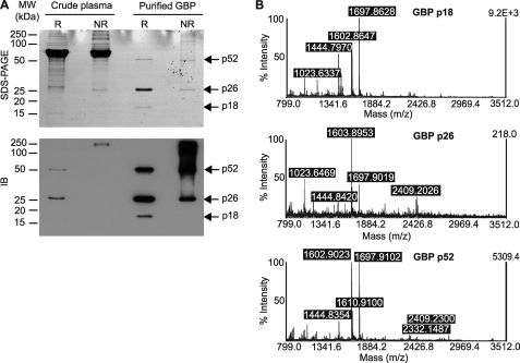
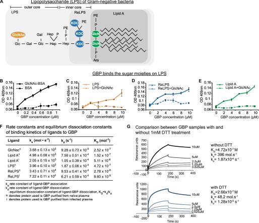
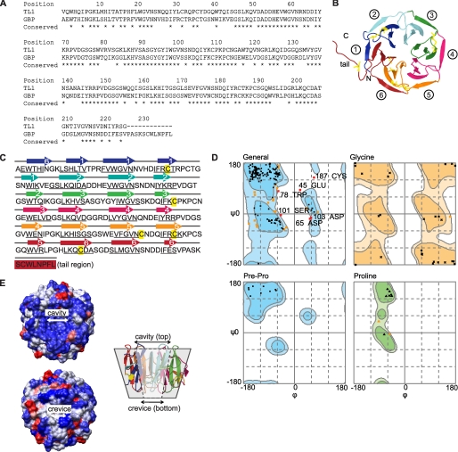
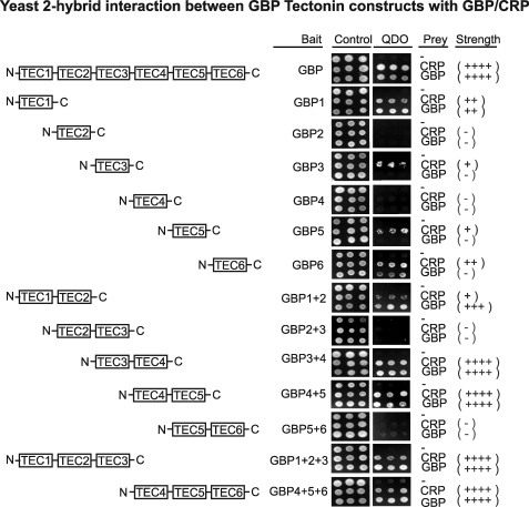
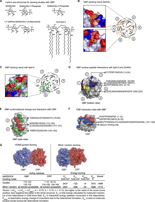
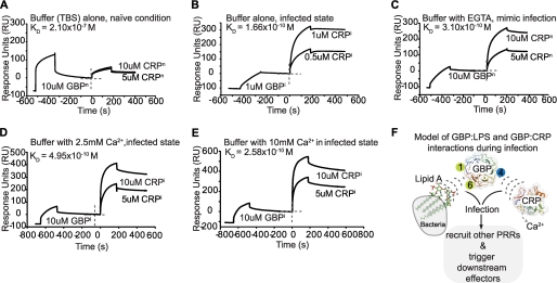
Beta-propeller proteins function in catalysis, protein-protein interaction, cell cycle regulation, and innate immunity. The galactose-binding protein (GBP) from the plasma of the horseshoe crab, Carcinoscorpius rotundicauda, is a beta-propeller protein that functions in antimicrobial defense. Studies have shown that upon binding to Gram-negative bacterial lipopolysaccharide (LPS), GBP interacts with C-reactive protein (CRP) to form a pathogen-recognition complex, which helps to eliminate invading microbes. However, the molecular basis of interactions between GBP and LPS and how it interplays with CRP remain largely unknown. By homology modeling, we showed that GBP contains six beta-propeller/Tectonin domains. Ligand docking indicated that Tectonin domains 6 to 1 likely contain the LPS binding sites. Protein-protein interaction studies demonstrated that Tectonin domain 4 interacts most strongly with CRP. Hydrogen-deuterium exchange mass spectrometry mapped distinct sites of GBP that interact with LPS and with CRP, consistent with in silico predictions. Furthermore, infection condition (lowered Ca(2+) level) increases GBP-CRP affinity by 1000-fold. Resupplementing the system with a physiological level of Ca(2+) did not reverse the protein-protein affinity to the basal state, suggesting that the infection-induced complex had undergone irreversible conformational change. We propose that GBP serves as a bridging molecule, participating in molecular interactions, GBP-LPS and GBP-CRP, to form a stable pathogen-recognition complex. The interaction interfaces in these two partners suggest that Tectonin domains can differentiate self/nonself, crucial to frontline defense against infection. In addition, GBP shares architectural and functional homologies to a human protein, hTectonin, suggesting its evolutionarily conservation for approximately 500 million years, from horseshoe crab to human.
Figures
Similar articles
-
A novel human tectonin protein with multivalent beta-propeller folds interacts with ficolin and binds bacterial LPS.PLoS One. 2009 Jul 16;4(7):e6260. doi: 10.1371/journal.pone.0006260. PLoS One. 2009. PMID: 19606221 Free PMC article.
-
C-reactive protein collaborates with plasma lectins to boost immune response against bacteria.EMBO J. 2007 Jul 25;26(14):3431-40. doi: 10.1038/sj.emboj.7601762. Epub 2007 Jun 21. EMBO J. 2007. PMID: 17581635 Free PMC article.
-
A female-specific pentraxin, CrOctin, bridges pattern recognition receptors to bacterial phosphoethanolamine.Eur J Immunol. 2007 Dec;37(12):3477-88. doi: 10.1002/eji.200737078. Eur J Immunol. 2007. PMID: 17979155
-
Molecular basis of non-self recognition by the horseshoe crab tachylectins.Biochim Biophys Acta. 2002 Sep 19;1572(2-3):414-21. doi: 10.1016/s0304-4165(02)00322-7. Biochim Biophys Acta. 2002. PMID: 12223283 Review.
-
Clotting and immune defense in Limulidae.Prog Mol Subcell Biol. 1996;15:154-89. doi: 10.1007/978-3-642-79735-4_8. Prog Mol Subcell Biol. 1996. PMID: 8963461 Review.
Cited by
-
Hydrogen/deuterium exchange mass spectrometry and site-directed disulfide cross-linking suggest an important dynamic interface between the two lysostaphin domains.Antimicrob Agents Chemother. 2013 Apr;57(4):1872-81. doi: 10.1128/AAC.02348-12. Epub 2013 Feb 4. Antimicrob Agents Chemother. 2013. PMID: 23380729 Free PMC article.
-
A review on host-pathogen interactions: classification and prediction.Eur J Clin Microbiol Infect Dis. 2016 Oct;35(10):1581-99. doi: 10.1007/s10096-016-2716-7. Epub 2016 Jul 29. Eur J Clin Microbiol Infect Dis. 2016. PMID: 27470504 Review.
-
Mapping the protein-protein interface between a toxin and its cognate antitoxin from the bacterial pathogen Streptococcus pyogenes.Biochemistry. 2011 May 17;50(19):4038-45. doi: 10.1021/bi200244k. Epub 2011 Apr 19. Biochemistry. 2011. PMID: 21466233 Free PMC article.
-
Structure-Function Relationships of C-Reactive Protein in Bacterial Infection.Front Immunol. 2019 Feb 26;10:166. doi: 10.3389/fimmu.2019.00166. eCollection 2019. Front Immunol. 2019. PMID: 30863393 Free PMC article. Review.
-
Methylated glycans as conserved targets of animal and fungal innate defense.Proc Natl Acad Sci U S A. 2014 Jul 8;111(27):E2787-96. doi: 10.1073/pnas.1401176111. Epub 2014 May 30. Proc Natl Acad Sci U S A. 2014. PMID: 24879441 Free PMC article.
References
-
- Fülöp V., Jones D. T. (1999) Curr. Opin. Struct. Biol. 9, 715–721 - PubMed
-
- Jawad Z., Paoli M. (2002) Structure 10, 447–454 - PubMed
-
- Chen S. C., Yen C. H., Yeh M. S., Huang C. J., Liu T. Y. (2001) J. Biol. Chem. 276, 9631–9639 - PubMed
-
- Huh C. G., Aldrich J., Mottahedeh J., Kwon H., Johnson C., Marsh R. (1998) J. Biol. Chem. 273, 6565–6574 - PubMed
Publication types
MeSH terms
Substances
LinkOut - more resources
Full Text Sources
Research Materials
Miscellaneous
