. 2010 Apr;192(7):1975-87.
doi: 10.1128/JB.01474-09. Epub 2010 Jan 29.

Histidine kinase-mediated production and autoassembly of Porphyromonas gingivalis fimbriae

Affiliations

Histidine kinase-mediated production and autoassembly of Porphyromonas gingivalis fimbriae

Kiyoshi Nishikawa et al. J Bacteriol. 2010 Apr.

Abstract

Porphyromonas gingivalis, a Gram-negative oral anaerobe, is strongly associated with chronic adult periodontitis, and it utilizes FimA fimbriae to persistently colonize and evade host defenses in the periodontal crevice. The FimA-related gene cluster (the fim gene cluster) is positively regulated by the FimS-FimR two-component system. In this study, comparative analyses between fimbriate type strain ATCC 33277 and fimbria-deficient strain W83 revealed differences in their fimS loci, which encode FimS histidine kinase. Using a reciprocal gene exchange system, we established that FimS from W83 is malfunctional. Complementation analysis with chimeric fimS constructs revealed that W83 FimS has a defective kinase domain due to a truncated conserved G3 box motif that provides an ATP-binding pocket. The introduction of the functional fimS from 33277 restored the production, but not polymerization, of endogenous FimA subunits in W83. Further analyses with a fimA-exchanged W83 isogenic strain showed that even the fimbria-deficient W83 retains the ability to polymerize FimA from 33277, indicating the assembly of mature FimA by a primary structure-dependent mechanism. It also was shown that the substantial expression of 33277-type FimA fimbriae in the W83 derivative requires the introduction and expression of the functional 33277 fimS. These findings indicate that FimSR is the unique and universal regulatory system that activates the fim gene cluster in a fimA genotype-independent manner.


Figures
FIG. 1.
FIG. 1.
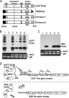
Reduced expression of fimSR and fim gene cluster in W83. (A) The structure of fimX-pgmA and fimA loci and the design of amplicons to be generated by RT-PCR. (B) Semiquantitative RT-PCR of fimA. After 22 PCR cycles with 300 ng of cDNA or 400 ng of RNA (negative control; data not shown) as a template, 10 μl out of 50 μl of each reaction mixture was run on a 1.5% agarose gel. (C) Semiquantitative RT-PCR (25 cycles) of the fimX-pgmA operon with reaction conditions similar to those described for panel B. (D) Schematic diagrams of fimSR loci in 33277 and W83. TM1 and TM2 indicate predicted transmembrane regions. In W83, the N-terminal cytoplasmic domain-coding region, derived from the 385-kbp insertional fragment, is depicted in black. Amplicons to be generated by RT-PCR also are indicated. (E) Semiquantitative RT-PCR of fimSR with 300 ng cDNA or 400 ng RNA (negative control; data not shown) as a template. After 25 cycles of the PCR, 10 μl out of 50 μl of each reaction mixture was run on a 2% agarose gel. M, DNA ladder marker. (F) Production of FimR protein in 33277 (lane 1), the fimR mutant (lane 2, AG21-5), the fimS mutant (lane 3, AGFS1), and W83 (lane 4). Total protein samples from each strain, adjusted to equal cell numbers, were fractionated by SDS-PAGE, transferred to a PVDF membrane, and probed with anti-FimR antibody. (G) Relative quantitation of fimSR and fimA-related gene expression in strains W83 and ATCC 33277. Reaction mixtures containing 100 ng of cDNA from either W83 or 33277 total RNA were subjected to real-time PCR as described in Materials and Methods. The expression of target genes was normalized to the endogenous 16S rRNA in each strain. The results are presented as ratios of target mRNA levels in W83 to those in 33277 by the calculation of 2−ΔΔCT, as described in Materials and Methods and Table S1 in the supplemental material. The error bar indicates standard deviations.
FIG. 2.
FIG. 2.
Comparison of fimS transcriptional start sites (TS) from 33277 and W83 by 5′ RLM-RACE. (A) Alignments of 33277 (327) and W83 fimS sequences around the FimS N-terminal coding regions. Originally predicted amino acid-coding sequences are shown in uppercase letters, and sequences from the inserted fragment in W83 are shown in italics. Arrows indicate three gene-specific reverse primers used for 5′ RACE (Table 1). (B) Results of nested PCR of 5′ RACE. Reaction mixtures were separated on a 3.5% agarose gel. The PCR products shown in lanes 1, 2, and 3 in both panels were amplified by using reverse primers GSP1, GSP2, and GSP3, respectively. The fragments indicated by arrowheads are TS-containing PCR products with the 29-bp 5′ adapter sequence. The products by GSP3 primer from both strains (lane 3) also were extracted and sequenced to determine TSs, which are boxed in Fig. 2A (A−18 for 33277 and G35 for W83).
FIG. 3.
FIG. 3.
Mutations found in the kinase domain-coding region of W83 fimS. (A) Two regions in the kinase domain of W83 FimS differ from that of 33277 FimS. 1, a single-amino-acid substitution from isoleucine to lysine; 2, truncation of the C-terminal region containing the G3 box. (B) Alignments of fimS nucleotide and deduced amino acid sequences around the FimS kinase domain. 1, a missense mutation (ATA [I511] to AAA [K518]) is located upstream of the N box (NLLSNA) coding region; 2, an insertion of A (indicated by an arrow) is found downstream of the G2 box (GSGLGL) coding region of W83. This insertion caused a frameshift mutation and truncated the C-terminal coding region that contains the predicted G3 box (GKGSVF). The VDD motif in FimR is conserved in the receiver domains of response regulators. (C) Alignments of amino acid sequences around the distinct regions, 1 and 2, of 33277 and W83 FimS (Pg327S and PgW83S, respectively) with histidine kinases from other Bacteroidetes. Bv, Bacteroides vulgatus ATCC 8482; Bt, Bacteroides thetaiotaomicron VPI-5482; and Bf, Bacteroides fragilis NCTC9343. Numbers followed by Bv, Bt, and Bf represent gene locus numbers of histidine kinases in each organism. Note that the predicted G3 box (G+GS+F) in region 2 is highly conserved beyond species, whereas the amino acid indicated by an asterisk in region 1 is not.
FIG. 4.
FIG. 4.
Phenotype of fimS-exchanged strains. (A) CBB-stained SDS-12.5% PAGE gel (upper) and Western blot (lower) with anti-type I FimA antibody. Total protein samples were denatured at 80°C for 20 min before SDS-PAGE. Lane 1, 33277; 2, AGFS1; 3, AGFS1(pTCBex); 4, AGFS1(pTCBex33277fimS); 5, AGFS1(pTCBexW83fimS); 6, W83; 7, WFS1; 8, WFS1(pTCBex); 9, WFS1(pTCBex33277fimS); 10, WFS1(pTCBexW83fimS). The CBB-stained single band of W83 FimA in lane 9 is indicated by a thin arrow. A thick arrow indicates the signals from 41-kDa monomers. (B) CBB-stained SDS-12.5% PAGE gel (upper) and Western blot with anti-W83 FimA antibody (lower). Total protein samples were denatured at 80°C for 20 min before being subjected to SDS-PAGE. Lane 1, W83; 2, W83(pTCBex); 3, W83(pTCBex33277fimS); 4, W83(pTCBexW83fimS); 5, WFS1; 6, WFS1(pTCBex); 7, WFS1(pTCBex33277fimS); 8, WFS1(pTCBexW83fimS); 9, 33277. Stained bands corresponding to W83 FimA in lanes 3 and 7 are indicated by an arrow. (C) RT-PCR confirming the expression of fimS in the complemented fimS knockout mutants. The 500-bp fimS cDNAs were amplified with the primer set shown in Fig. 1D and Table 1. After 25 cycles of PCR, an equal volume of each reaction mixture was fractionated by 3% AGE. The glucose kinase gene (glk; 415 bp) was coamplified in each reaction as an internal control. Lane 1, 33277; 2, AGFS1(pTCBex); 3, AGFS1(pTCBex33277fimS); 4, AGFS1(pTCBexW83fimS); 5, W83; 6, W83(pTCBex); 7, W83(pTCBex33277fimS); 8, W83(pTCBexW83fimS); 9, WFS1(pTCBex); 10, WFS1(pTCBex33277fimS); 11, WFS1(pTCBexW83fimS).
FIG. 5.
FIG. 5.
Truncation of G3 box is responsible for functional deficiency of W83 FimS. (A) Design of W83-33277 fimS chimeras. Chimera-1 consists of an N-terminal cytoplasmic domain-coding region from W83 and C-terminal kinase domain-coding region from 33277 possessing the G3 box. Chimera-2 is a derivative of Chimera-1 with the same missense mutation, AAA, as that in W83 fimS. (B) Upper panel, a Western blot with anti-type I FimA antibodies showing induced levels of 33277 FimA by the complementation of the fimS knockout ATCC 33277 with fimS variants. Samples were heated at 80°C for 20 min prior to SDS-12.5% PAGE to observe the polymerization of FimA. Lower panel, expression of introduced fimS revealed by RT-PCR. The coamplified glucose kinase gene (glk) was an internal control. Lane 1, 33277; 2, AGFS1(pTCBex); 3, AGFS1(pTCBex33277fimS); 4, AGFS1(pTCBexW83fimS); 5, AGFS1(pTCBexChimera-1); 6, AGFS1(pTCBexChimera-2). (C) Upper panel, a Western blot with anti-type IV FimA antibodies showing induced levels of W83 FimA by the complementation of the fimS knockout W83 with fimS variants. Samples were boiled for 20 min before SDS-PAGE (12.5%) to maximize signal intensities for detection. Lower panel, expression of introduced fimS revealed by RT-PCR. Lane 1, WFS1(pTCBex); 2, WFS1(pTCBex33277fimS); 3, WFS1(pTCBexW83fimS); 4, WFS1(pTCBexChimera-1); 5, WFS1(pTCBexChimera-2). (D) Summarizing model. The G3 box, in addition to G1 and G2 boxes, in the C terminus of the 33277 FimS kinase domain forms a pocket to bind ATP, which autophosphorylates the histidine residue of the H box. The phosphoryl group then is transferred to the cognate response regulator, FimR, to activate its function as a positive regulator of the fim gene cluster. The truncation of the G3 box in W83 FimS renders it defective in ATP capture, leading to the disruption of the subsequent phosphorelay.
FIG. 6.
FIG. 6.
33277 FimS-induced W83 FimA is a mature polypeptide and is localized on the cell surface. (A) Whole cells were heat treated at 37°C (lanes 1 and 5), 50°C (lanes 2 and 6), 80°C (lanes 3 and 7), and 100°C (lanes 4 and 8) and fractionated by SDS-PAGE, and Western blots were probed with strain-specific anti-FimA antibody. Left, ATCC 33277; right, WFS1(pTCBex33277fimS). (B) Localization of W83 FimA in WFS1. WFS1(pTCBex33277fimS) cells were fractioned and analyzed by Western blotting. Lane 1, culture supernatant; 2, whole cell; 3, cytoplasm/periplasm; 4, envelope; 5, inner membrane; 6, outer membrane. The upper panel shows a membrane probed with anti-W83 FimA antibody. The same samples were transferred onto an equivalent membrane and probed with anti-recombinant PgmA antibody to validate the fractionation (lower panel). (C) Alignment of N-terminal sequences of 33277 and W83 FimA proteins. Boxed sequences (MTAC) are the lipoprotein sorting signals, and lysine (K) and arginine (R) residues of gingipain cleavage sites are highlighted in boldface and a larger size (35). An arrow indicates the first residue (A47) of mature 33277 FimA. The first 10 residues of W83 FimA from outer membrane fraction, determined by an N-terminal peptide sequence analysis, are underlined and highlighted in boldface. (D) Schematic representations showing the distinct phenotype of FimA fimbriae in the P. gingivalis strains studied. Strain W83 lacks FimA fimbriae due to the deficiency in the positive regulation of the fim gene cluster, as depicted in Fig. 5D. In W83 or the fimS knockout derivative (WFS1) expressing 33277 FimS in trans, W83 FimA propeptides are transported to the cell surface and cleaved by RGP to mature monomers. The monomers remain at the cell surface and are not polymerized.
FIG. 7.
FIG. 7.
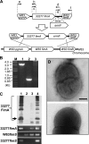
W83 retains the ability to assemble 33277-type FimA fimbriae. (A) Construction of fimA-exchanged W83 variant W83fA327. A chimeric DNA fragment comprised of the C-terminal coding region of W83 pgmA, full-length 33277 fimA, the N-terminal coding region of W83 fimB, and intergenic sequences from W83 was generated by a PCR-based overlap extension, as described in Materials and Methods. After the insertion of an ermF cassette, the final construct was introduced into W83 by electroporation. (B) Confirmation of allelic exchange by genomic DNA PCR. Lane 1, amplicons with primers a and f (1,968 bp + 1,095 bp of ermF = 3,063 bp; this amplicon also was sequenced); lane 2, amplicons with specific primers for 33277 fimA (700 bp); lane 3, no 700-bp amplicons with W83 fimA-specific primers. (C) Complementation of the fimA-exchanged W83 with the functional 33277 fimS gene is sufficient for the assembly of FimA fimbriae. The upper panel shows a Western blot of 80°C heat-treated whole-cell proteins from 33277 (lane 1), W83 (lane 2), W83fA327 harboring pTCBex (lane 3), and W83fA327 harboring pTCBex33277fimS (lane 4) probed with anti-33277 FimA antibody. Ladder signals are from polymerized FimA. An arrow indicates the signals from 41-kDa monomers. Lower panels represent the expression profile of each strain by RT-PCR. (D) Morphological observation of fimbrial assembly by transmission electron microscopy. The upper panel shows fimbriate W83fA327 harboring pTCBex33277fimS. The lower panel shows a fimbria-less W83 derivative expressing W83 fimA and 33277 fimS (i.e., strain WFS1 harboring pTCBex33277fimS). Scale bar, 200 nm.

Similar articles

Cited by

References

    1. Akiyama, Y., K. Onizuka, T. Noguchi, and M. Ando. 1998. Parallel protein information analysis (PAPIA) system running on a 64-node PC cluster, p. 131-140. In Proc. 9th Genome Informatics Workshop (GIW'98), Universal Academy Press, Tokyo, Japan. - PubMed
    1. Amano, A., I. Nakagawa, K. Kataoka, I. Morisaki, and S. Hamada. 1999. Distribution of Porphyromonas gingivalis strains with fimA genotypes in periodontal patients. J. Clin. Microbiol. 37:1426-1430. - PMC - PubMed
    1. Belton, C. M., K. T. Izutsu, P. C. Goodwin, Y. Park, and R. J. Lamont. 1999. Fluorescence image analysis of the association between Porphyromonas gingivalis and gingival epithelial cells. Cell. Microbiol. 1:215-223. - PubMed
    1. Chen, T., Y. Hosogi, K. Nishikawa, K. Abby, R. D. Fleischmann, J. Walling, and M. J. Duncan. 2004. Comparative whole genome analysis of virulent and avirulent strains of Porphyromonas gingivalis. J. Bacteriol. 186:5473-5479. - PMC - PubMed
    1. Das, A. K., P. T. W. Cohen, and D. Barford. 1998. The structure of the tetratricopeptide repeats of protein phosphatase 5: implications for TPR-mediated protein-protein interactions. EMBO J. 17:1192-1199. - PMC - PubMed

Publication types

MeSH terms

LinkOut - more resources