Comparative Study
. 2010 Mar;34(3):327-40.
doi: 10.1097/PAS.0b013e3181cd3aeb.

B-cell lymphomas with concurrent IGH-BCL2 and MYC rearrangements are aggressive neoplasms with clinical and pathologic features distinct from Burkitt lymphoma and diffuse large B-cell lymphoma

Affiliations
Comparative Study

B-cell lymphomas with concurrent IGH-BCL2 and MYC rearrangements are aggressive neoplasms with clinical and pathologic features distinct from Burkitt lymphoma and diffuse large B-cell lymphoma

Matija Snuderl et al. Am J Surg Pathol. 2010 Mar.

Abstract

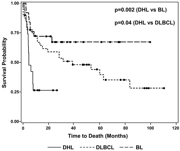
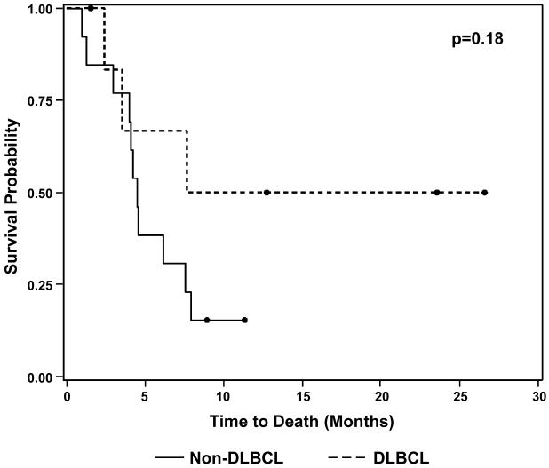
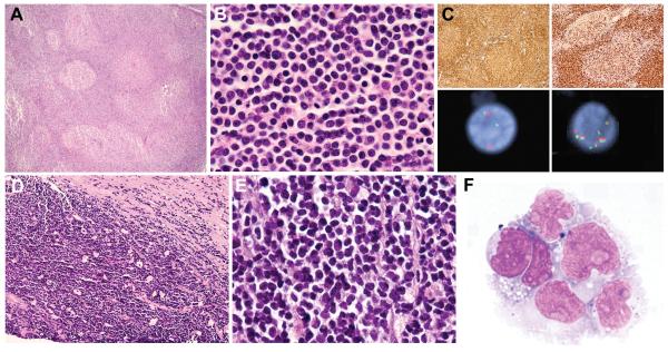
B-cell lymphomas with concurrent IGH-BCL2 and MYC rearrangements, also known as "double-hit" lymphomas (DHL), are rare neoplasms characterized by highly aggressive clinical behavior, complex karyotypes, and a spectrum of pathologic features overlapping with Burkitt lymphoma (BL), diffuse large B-cell lymphoma (DLBCL) and B-lymphoblastic lymphoma/leukemia (B-LBL). The clinical and pathologic spectrum of this rare entity, including comparison to other high-grade B-cell neoplasms, has not been well defined. We conducted a retrospective analysis of clinical and pathologic features of 20 cases of DHL seen at our institution during a 5-year period. In addition, we carried out case-control comparisons of DHL with BL and International Prognostic Index (IPI)-matched DLBCL. The 11 men and 9 women had a median age of 63.5 years (range 32 to 91). Six patients had a history of grade 1 to 2 follicular lymphoma; review of the prior biopsy specimens in 2 of 5 cases revealed blastoid morphology. Eighteen patients had Ann Arbor stage 3 or 4 disease and all had elevated serum lactate dehydrogenase (LDH) levels at presentation. Extranodal disease was present in 17/20 (85%), bone marrow involvement in 10/17 (59%) and central nervous system (CNS) disease in 5/11 (45%). Nineteen patients were treated with combination chemotherapy, of whom 18 received rituximab and 14 received CNS-directed therapy. Fourteen patients (70%) died within 8 months of diagnosis. Median overall survival in the DHL group (4.5 mo) was inferior to both BL (P=0.002) and IPI-matched DLBCL (P=0.04) control patients. Twelve DHL cases (60%) were classified as B-cell lymphoma, unclassifiable, with features intermediate between DLBCL and BL, 7 cases (35%) as DLBCL, not otherwise specified, and 1 case as B-LBL. Distinguishing features from BL included expression of Bcl2 (P<0.0001), Mum1/IRF4 (P=0.006), Ki-67 <95% (P<0.0001), and absence of EBV-EBER (P=0.006). DHL commonly contained the t(8;22) rather than the t(8;14) seen in most BL controls (P=0.001), and exhibited a higher number of chromosomal aberrations (P=0.0009). DHL is a high-grade B-cell neoplasm with a poor prognosis, resistance to multiagent chemotherapy, and clinical and pathologic features distinct from other high-grade B-cell neoplasms. Familiarity with the morphologic and immunophenotypic spectrum of DHL is important in directing testing to detect concurrent IGH-BCL2 and MYC rearrangements when a karyotype is unavailable. The aggressive clinical behavior and combination of genetic abnormalities seen in these cases may warrant categorization as a separate entity in future classifications and call for novel therapeutic approaches.


Figures

Figure 1
Figure 1
Kaplan-Meier Overall Survival Distributions for Double-Hit Lymphoma, Burkitt Lymphoma and IPI-Matched Diffuse Large B-cell Lymphoma Patients Black circles denote patients who were alive at the time of last follow-up. DHL: double-hit lymphoma, BL: Burkitt lymphoma, DLBCL: diffuse large B-cell lymphoma.
Figure 2
Figure 2
B-Lymphoblastic Leukemia with IGH-BCL2 and MYC Rearrangements The bone marrow core biopsy from case 20 showed medium-sized cells with round nuclei and finely dispersed chromatin in a background of extensive cellular necrosis (A). The peripheral blood contained a significant population of blasts with round to irregular nuclei, prominent nucleoli, dispersed chromatin and deeply basophilic cytoplasm (B-C). Immunophenotyping of the circulating leukemic cells by flow cytometry showed a population of CD19+, CD10+, terminal deoxynucleotidyl transferase (TdT)+ B lymphoblasts (D) that were CD45dim+ and negative for CD20 and surface light chain (not shown).
Figure 3
Figure 3
Morphologic Spectrum of Double-Hit Lymphoma Five double-hit lymphoma cases classified as B-cell lymphoma, unclassifiable (BCLU), with features intermediate between diffuse large B-cell lymphoma (DLBCL) and Burkitt lymphoma (BL) showed Burkitt-like morphological features, with a prominent starry-sky growth pattern at low magnification (A: cervical lymph node, case 3) and a population of monomorphous medium-sized cells with round to slightly irregular nuclei, finely clumped chromatin and multiple small nucleoli at high magnification (B: axillary lymph node, case 2). Four BCLU cases were morphologically intermediate between BL and DLBCL with greater cytomorphologic variation, including medium-sized to slightly larger cells containing conspicuous nuclear irregularity and single prominent central nucleoli (C: abdominal wall subcutaneous soft tissue, case 9). Three BCLU cases exhibited blastoid cytologic features, with small cells containing finely dispersed chromatin and inconspicuous nucleoli (D: testicle, case 10). Cases classified as diffuse large B-cell lymphoma, not otherwise specified (DLBCL-NOS) contained predominantly large atypical cells with oval to irregular nuclei, including some with prominent central nucleoli (E: bone marrow biopsy, case 19). The corresponding aspirate smear contained a population of large cells with irregular nuclei, prominent nucleoli, and deeply basophilic cytoplasm with conspicuous vacuolation (F).
Figure 4
Figure 4
Immunophenotypic, Cytogenetic and Molecular Genetic Features of Double-Hit Lymphoma Two examples of double-hit lymphoma (DHL) demonstrating a Ki-67 proliferation index (PI) of <95%: a retroperitoneal lymph node biopsy from case 5, classified as B-cell lymphoma, unclassifiable, with features intermediate between diffuse large B-cell lymphoma and Burkitt lymphoma (BCLU), with an overall Ki-67 PI of 60% (A); and an abdominal lymph node biopsy from case 14, classified as diffuse large B-cell lymphoma, not otherwise specified (DBLCL-NOS), with an overall Ki-67 PI of 80% (B). The vast majority of DHL cases expressed Bcl2 by immunohistochemistry for the commonly used antibody, clone 124 (C: stomach polyp, case 1). All DHL cases were of germinal center origin, expressing CD10, Bcl6, or both, as in this cervical lymph node biopsy from case 12 (D: CD10, E: Bcl6). Mum1 was expressed in 8/19 DHL cases, including this axillary lymph node biopsy from case 2 (F). Bone marrow cytogenetic analysis from case 4 showed a complex karyotype (G, arrows), as did all DHL cases. In addition to the t(8;22) and t(14;18), this case contained additional unknown material in the 3q2?7 region (G, arrow indicating chromosome 3). Subsequent fluorescence in situ hybridization (FISH) analysis confirmed a rearrangement involving BCL6 at 3q27 (not shown). Interphase FISH analysis of paraffin-embedded stomach polyp tissue from case 1 confirmed both a MYC rearrangement (H: dual-color, split-apart probe) and an IGH-BCL2 fusion (I: dual-color, dual fusion probe). The FISH patterns with both probes were consistent with unbalanced rearrangements (H-I). This patient’s prior low-grade follicular lymphoma lacked both MYC and BCL2 rearrangements (not shown), suggesting that both translocations were acquired at the time of DHL transformation.
Figure 5
Figure 5
Differences in Kaplan-Meier Overall Survival Distributions for Double-Hit Lymphoma Patients by 2008 WHO Classification Black circles denote patients who were alive at the time of last follow-up. DLBCL: diffuse large B-cell lymphoma, not otherwise specified.
Figure 6
Figure 6
Pre-Existing Low-Grade Follicular Lymphomas with Unusual Features Examination of prior low-grade follicular lymphomas in 2 patients revealed findings unusual for this diagnosis. In case 10 with testicular double-hit lymphoma (DHL; illustrated in Fig. 3D), review of a supraclavicular lymph node biopsy from 2 years earlier showed architectural effacement by a follicular proliferation of B cells (A). Cells within and outside of follicles were small with blastoid features, including round nuclei, evenly dispersed chromatin and absent nucleoli (B); typical centrocytes were not identified. Both follicular and extrafollicular B cells were strongly CD20+, CD10+ and Bcl2+ (C, upper left). The follicular component weakly co-expressed Bcl6, but showed absent follicular dendritic cell staining (not shown). Ki-67 stain showed an unusually high proliferation index (PI) of approximately 40% within follicles and 90% outside of follicles (C, upper right). Fluorescence in situ hybridization (FISH) analysis with dual color break apart probes revealed a MYC rearrangement (C, lower left) and 8 copies of BCL2 (C, lower right), the same genetic abnormalities seen in the subsequent DHL. In case 11, core needle biopsies of a retroperitoneal mass showed a CD10+, Bcl6+, Bcl2+ B-cell neoplasm with a diffuse growth pattern (absent staining for follicular dendritic cell antigens), a focal starry-sky pattern (D) and patchy necrosis. The cells were small to medium-sized; some were centrocyte-like, but the majority had oval nuclei with finely dispersed chromatin and absent nucleoli (E), prompting a diagnosis of diffuse grade 1-2 FL with blastoid features. Tissue from the initial biopsies was not available for cytogenetics or FISH analysis. Six months later, the patient relapsed with a CD10+ B-cell lymphoma involving the central nervous system (F), confirmed by FISH to have an IGH-BCL2 fusion as well as a MYC rearrangement (FISH analysis not shown).

Similar articles

Cited by

References

    1. The International Non-Hodgkin’s Lymphoma Prognostic Factors Project A predictive model for aggressive non-Hodgkin’s lymphoma. N Engl J Med. 1993;329:987–994. - PubMed
    1. Boerma EG, Siebert R, Kluin PM, et al. Translocations involving 8q24 in Burkitt lymphoma and other malignant lymphomas: a historical review of cytogenetics in the light of today’s knowledge. Leukemia. 2009;23:225–234. - PubMed
    1. Brito-Babapulle V, Crawford A, Khokhar T, et al. Translocations t(14;18) and t(8;14) with rearranged bcl-2 and c-myc in a case presenting as B-ALL (L3) Leukemia. 1991;5:83–87. - PubMed
    1. Cogliatti SB, Novak U, Henz S, et al. Diagnosis of Burkitt lymphoma in due time: a practical approach. Br J Haematol. 2006;134:294–301. - PubMed
    1. Cox DR. Partial likelihood. Biometrika. 1975;62:277–288.

Publication types

MeSH terms

Substances