. 2010 Feb;28(2):167-71.
doi: 10.1038/nbt.1604. Epub 2010 Jan 31.

Real-time imaging of hepatitis C virus infection using a fluorescent cell-based reporter system

Affiliations

Real-time imaging of hepatitis C virus infection using a fluorescent cell-based reporter system

Christopher T Jones et al. Nat Biotechnol. 2010 Feb.

Abstract

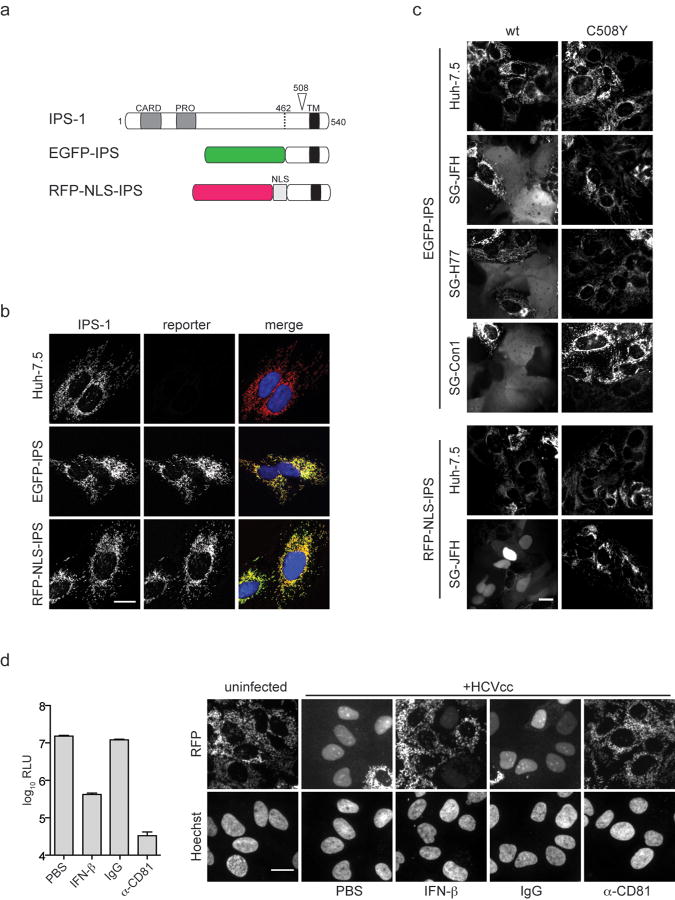
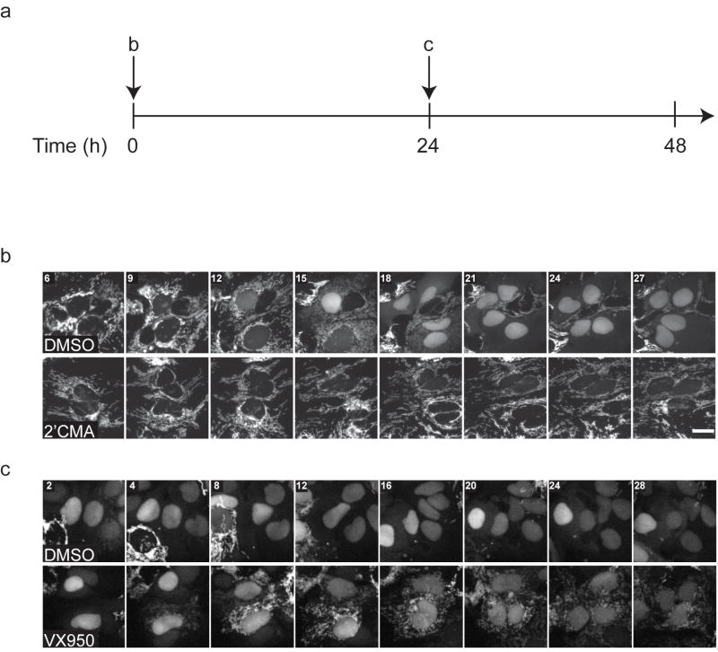
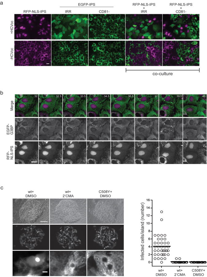
Hepatitis C virus (HCV), which infects 2-3% of the world population, is a causative agent of chronic hepatitis and the leading indication for liver transplantation. The ability to propagate HCV in cell culture (HCVcc) is a relatively recent breakthrough and a key tool in the quest for specific antiviral therapeutics. Monitoring HCV infection in culture generally involves bulk population assays, use of genetically modified viruses and/or terminal processing of potentially precious samples. Here we develop a cell-based fluorescent reporter system that allows sensitive distinction of individual HCV-infected cells in live or fixed samples. We demonstrate use of this technology for several previously intractable applications, including live-cell imaging of viral propagation and host response, as well as visualizing infection of primary hepatocyte cultures. Integration of this reporter with modern image-based analysis methods could open new doors for HCV research.


Conflict of interest statement

Competing Financial Interests The authors declare the following conflicts of interest, which are managed under University policy: C.M.R. has equity in Apath, LLC, which holds commercial licenses for the Huh-7.5 cell line and the HCVcc cell culture system. S.R.K and S.N.B have equity in Hepregen, Corporation, which holds commercial licenses from the Massachusetts Institute of Technology for micropatterned co-cultures (MPCCs) and other related microscale liver technologies.

Figures

Figure 1
Figure 1. An IPS-1-based reporter system for detection of HCV infection
(a) Schematic of IPS-1 and derivative reporter constructs. The Caspase Recruitment Domain (CARD) and proline rich (PRO) domains of IPS-1, also known as MAVS, VISA, or Cardif, are indicated. The HCV NS3-4A protease cleaves IPS-1 at C508 (arrow). The C-terminal transmembrane domain (TM) directs IPS-1 to the outer membrane of mitochondria. EGFP-IPS encodes EGFP fused to residues 462-540 of IPS-1. RFP-NLS-IPS encodes a red fluorescent protein (mCherry or TagRFP) and an SV40 nuclear localization signal (NLS, PKKKRKVG) fused to residues 462-540 of IPS-1. (b) EGFP-IPS and RFP-NLS-IPS localize to mitochondria in Huh-7.5 cells. Native IPS-1, detected by immunofluorescent staining (IPS-1), as well as EGFP or RFP autofluorescence (reporter) were visualized in untransduced (Huh-7.5) or transduced (EGFP-IPS or RFP-NLS-IPS) cells by confocal microscopy. Merge images also depict Hoechst nuclear dye (blue). (c) EGFP-IPS and RFP-NLS-IPS relocalize in response to HCV replication. Huh-7.5 cell lines harboring subgenomic (SG) neomycin-selectable replicons were transduced with lentiviruses expressing wild type (wt) or mutant (C508Y) EGFP-IPS or RFP-NSL-IPS. H77, genotype 1a; Con1, genotype 1b; JFH-1, genotype 2a. Wide-field fluorescence images of unfixed cells are shown. (d) RFP-NLS-IPS relocalizes in HCV infected cells. Huh-7.5 cells expressing RFP-NLS-IPS were infected with secreted Gaussia luciferase HCVcc reporter virus, Jc1FLAG2(p7-nsGluc2A), in the presence of phosphate buffered saline (PBS), IFN-β, blocking antibody (α-CD81) or isotype control (IgG). Luciferase activity in the culture supernatants (left) and reporter (RFP) or nuclear dye (Hoechst) fluorescence (right) were monitored at 48 h post-infection. Wide-field fluorescence images of fixed cells are shown. Scale bars, 20 μm. RLU, relative light units.
Figure 2
Figure 2. Time-lapse live-cell imaging of HCVcc infection
(a) Schematic of live-cell imaging time course. Huh-7.5 cells stably expressing RFP-NLS-IPS and a mitochondrially targeted EGFP-cytochrome c oxidase subunit VIII fusion protein (mito-EGFP) were infected with HCVcc reporter virus, Jc1FLAG2(p7-nsGluc2A). (b) Cells were infected in the presence of DMSO or HCV RNA-dependent RNA polymerase inhibitor 2’CMA. (c) Cells were infected for 24 h prior to removal of the inoculum and addition of imaging medium containing DMSO or the NS3-4A protease inhibitor VX950. Images were captured every 30 min starting at 6 h (b) or 24.5 h (c) post-infection. RFP fluorescence is shown in grayscale. Time (h) from the start of infection (b) or drug addition (c) are indicated. Scale bar, 20 μm. See Supplementary Videos 1a-d for full time course.
Figure 3
Figure 3. Use of the IPS-1-based reporter to expand HCVcc culture systems
(a) Co-culture of spectrally distinct HCV reporter cell lines for visualizing CD81-independent infection. Wide-field fluorescence images of unfixed mono- and co-cultures of Huh-7.5 cell lines expressing RFP-NLS-IPS or EGFP-IPS in the presence (+HCVcc) or absence (-HCVcc) of J6/JFH clone 2. EGFP-IPS cells stably express shRNA targeting CD81 (CD81-) or an irrelevant sequence (IRR). Monocultures were infected for 72 h prior to imaging. In co-culture experiments, RFP-NLS-IPS cells were infected with HCVcc for 36 h prior to mixing with uninfected EGFP-IPS/IRR or EGFP-IPS/CD81- cells. Co-cultures were incubated for an additional 48 h prior to imaging. (b) Multiplexing the HCV reporter with a marker of the stress response. An Huh-7 cell line expressing RFP-NLS-IPS and an EGFP-tagged stress granule marker (EGFP-G3BP) was infected with Jc1FLAG2(p7-nsGluc2A). Live-cell imaging was initiated at 6 h post-infection with images captured every 30 min. Montage shows selected time points beginning at 32 h post-infection; times (h) from the start of infection are indicated. See Supplementary Videos 2a-c. (c) Visualization of HCVcc infection in primary hepatocytes. Primary human hepatocytes maintained as micropatterned co-cultures (MPCC) were transduced with lentiviruses expressing wild type (wt) or mutant (C508Y) RFP-NLS-IPS. At 24 h post-transduction, MPCC were infected with Jc1FLAG2(p7-nsGluc2A). After 12 h, virus was removed and MPCC medium containing DMSO or 2’CMA was added. Unfixed MPCCs were imaged by wide-field fluorescence microscopy at 48 h post-infection. Representative phase contrast (top row) and corresponding RFP fluorescence images (middle row) are shown. Enlarged fluorescence images (bottom row) correspond to area denoted by white dotted box (middle row). The number of cells per MPCC island exhibiting nuclear RFP at 48 h post-infection is plotted for each condition. For wt RFP-NLS-IPS+DMSO n=40; wt RFP-NLS-IPS+2’CMA n=30; C508Y RFP-NLS-IPS+DMSO n=35. Bar, mean number of positive cells/island. Scale bars, 20 μm (a and b), 200 μm (c, top panel), 10 μm (c, lower panel).

Comment in

References

    1. Shepard CW, Finelli L, Alter MJ. Global epidemiology of hepatitis C virus infection. Lancet Infect Dis. 2005;5:558–567. - PubMed
    1. Meylan E, et al. Cardif is an adaptor protein in the RIG-I antiviral pathway and is targeted by hepatitis C virus. Nature. 2005;437:1167–1172. - PubMed
    1. Foy E, et al. Control of antiviral defenses through hepatitis C virus disruption of retinoic acid-inducible gene-I signaling. Proc Natl Acad Sci U S A. 2005;102:2986–2991. - PMC - PubMed
    1. Li XD, Sun L, Seth RB, Pineda G, Chen ZJ. Hepatitis C virus protease NS3/4A cleaves mitochondrial antiviral signaling protein off the mitochondria to evade innate immunity. Proc Natl Acad Sci U S A. 2005;102:17717–17722. - PMC - PubMed
    1. Kawai T, et al. IPS-1, an adaptor triggering RIG-I- and Mda5-mediated type I interferon induction. Nat Immunol. 2005;6:981–988. - PubMed

Publication types

MeSH terms