. 2009 Aug;3(4):223-7.
doi: 10.2976/1.3185785. Epub 2009 Jul 24.

Traction forces during collective cell motion

Affiliations

Traction forces during collective cell motion

N S Gov. HFSP J. 2009 Aug.

Abstract

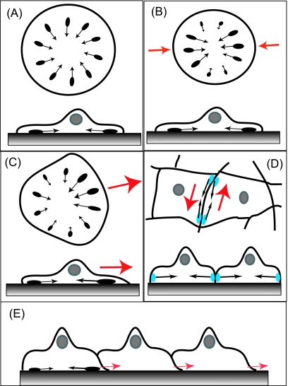
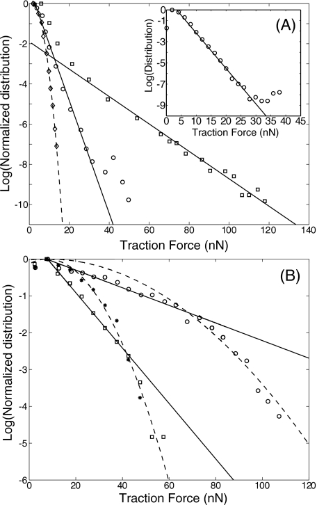
Collective motion of cell cultures is a process of great interest, as it occurs during morphogenesis, wound healing, and tumor metastasis. During these processes cell cultures move due to the traction forces induced by the individual cells on the surrounding matrix. A recent study [Trepat, et al. (2009). Nat. Phys. 5, 426-430] measured for the first time the traction forces driving collective cell migration and found that they arise throughout the cell culture. The leading 5-10 rows of cell do play a major role in directing the motion of the rest of the culture by having a distinct outwards traction. Fluctuations in the traction forces are an order of magnitude larger than the resultant directional traction at the culture edge and, furthermore, have an exponential distribution. Such exponential distributions are observed for the sizes of adhesion domains within cells, the traction forces produced by single cells, and even in nonbiological nonequilibrium systems, such as sheared granular materials. We discuss these observations and their implications for our understanding of cellular flows within a continuous culture.


Figures

Figure 1
Figure 1. Modes of traction forces produced by cells.
(A) Traction forces within a nonmotile cell are produced at the cell-substrate contacts (black ovals representing FA domains) due to the contractile forces produced by the actin-myosin cytoskeleton (black arrows). These forces are uniform around the cell, pointing inwards, and result in a vanishing net traction. (B) In a polarized nonmotile cell the traction forces have the form of a “force-dipole” (red arrows). (C) In a motile cell the traction forces are unbalanced, being larger at the leading edge (lamellipodia), and the net traction force leads to cell motility (red arrow). (D) In a multicellular culture traction forces arise at cell-cell contacts (blue ovals) and can lead to cells sliding past each other (red arrows), and to cells pulling toward each other. (E) Inside a continuous culture cells produce cell-substrate traction due to polarized lamellipodia (red arrows), which will naturally tend to polarize neighboring cells in the same direction.
Figure 2
Figure 2. Exponential distribution of traction forces produced by cells.
(A) Distribution of traction forces measured (Tymchenko et al., 2007) for endothelial (circles) and fibroblast (squares) cells on a silicon substrate. Dashed line is a fit to a Gaussian distribution for the measured background noise (diamonds), while solid lines give fits to exponential distributions. Inset: Distribution of traction forces measured (du Roure et al., 2005) at the edge of an expanding culture of MDCK cells (circles). (B) Distribution of traction forces measured (Ganz et al., 2006) for (i) C2 cells on N-cadherin-coated (stars) and fibronectin-coated (circles) micropillars, and for (ii) GT1–7 cell on N-cadherin-coated micropillars (squares). Dashed lines are fits to Gaussian distributions, while solid lines give fits to exponential distributions.

Comment on

  • Trepat, et al. (2009). Nat. Phys. 5, 426–430

Similar articles

Cited by

References

    1. Balaban, N Q, Schwarz, U S, Riveline, D, Goichberg, P, Tzur, G, Sabanay, I, Mahalu, D, Safran, S, Bershadsky, A, Addadi, L, and Geiger, B (2001). “Force and focal adhesion assembly: a close relationship studied using elastic micro-patterned substrates.” Nat. Cell Biol. NCBIFN 3, 466–472.10.1038/35074532 - DOI - PubMed
    1. Beningo, K A, Dembo, M, Kaverina, I, Small, J V, and Wang, YL (2001). “Nascent focal adhesions are responsible for the generation of strong propulsive forces in migrating fibroblasts.” J. Cell Biol. JCLBA3 153, 881–887.10.1083/jcb.153.4.881 - DOI - PMC - PubMed
    1. Beningo, K A, and Wang, Y L (2002). “Flexible substrata for the detection of cellular traction forces.” Trends Cell Biol. TCBIEK 12, 79–84.10.1016/S0962-8924(01)02205-X - DOI - PubMed
    1. Besser, A, and Safran, S A (2006). “Force-induced adsorption and anisotropic growth of focal adhesions.” Biophys. J. BIOJAU 90, 3469–3484.10.1529/biophysj.105.074377 - DOI - PMC - PubMed
    1. Butler, B, Gao, C, Mersich, A T, and Blystone, SD (2006). “Purified integrin adhesion complexes exhibit actin-polymerization activity.” Curr. Biol. CUBLE2 16, 242–251.10.1016/j.cub.2005.12.033 - DOI - PubMed

LinkOut - more resources