. 2009 Aug;3(4):240-54.
doi: 10.2976/1.3137602. Epub 2009 Jun 3.

Experimental and computational aspects of signaling mechanisms of spike-timing-dependent plasticity

Experimental and computational aspects of signaling mechanisms of spike-timing-dependent plasticity

Hidetoshi Urakubo et al. HFSP J. 2009 Aug.

Abstract

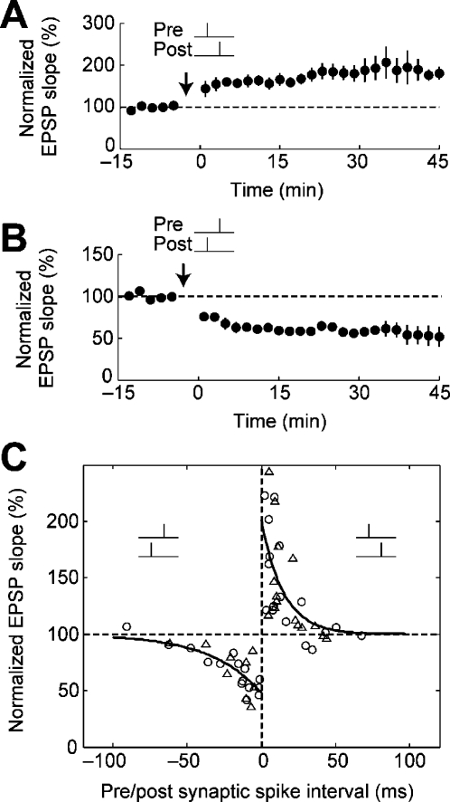
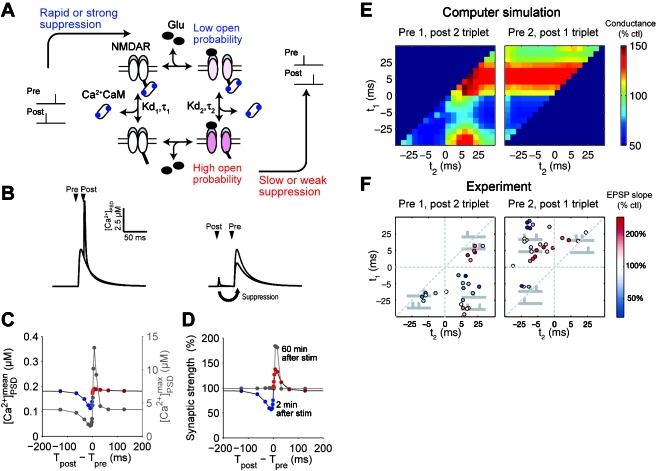
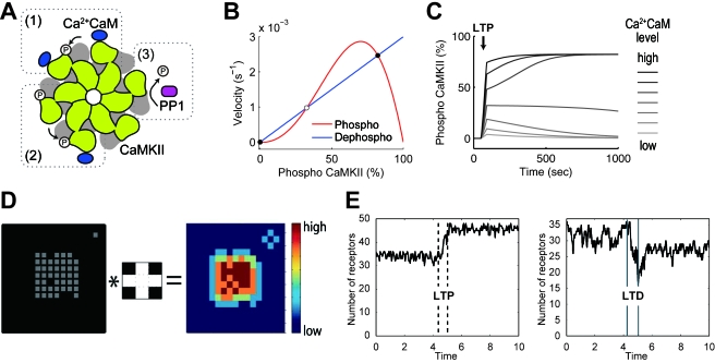
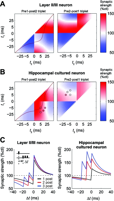
STDP (spike-timing-dependent synaptic plasticity) is thought to be a synaptic learning rule that embeds spike-timing information into a specific pattern of synaptic strengths in neuronal circuits, resulting in a memory. STDP consists of bidirectional long-term changes in synaptic strengths. This process includes long-term potentiation and long-term depression, which are dependent on the timing of presynaptic and postsynaptic spikings. In this review, we focus on computational aspects of signaling mechanisms that induce and maintain STDP as a key step toward the definition of a general synaptic learning rule. In addition, we discuss the temporal and spatial aspects of STDP, and the requirement of a homeostatic mechanism of STDP in vivo.


Figures

Figure 1
Figure 1. A canonical form of STDP.
Reprinted by permission, from Macmillan Publishers Ltd: Nature (Froemke and Dan, 2002), copyright (2002). (A) Repetitive pairings of prespiking→postspiking within a 20 ms interval (20 ms>TpostTpre>0 ms) at 0.2 Hz lead to persistent increase In synaptic strength (LTP). (B) Repetitive pairings of postspiking→prespiking within a 40 ms interval (−40 ms<TpostTpre<0 ms) at 0.2 Hz lead to persistent decrease in synaptic strength (LTD). (C) The critical timing window of STDP. Circles and triangles indicate experimental data (triangles: high Ca2+ and Mg2+ with bicuculline), and solid lines are exponential fits to the data. The data are taken from layer II∕III neurons in the visual cortex (Froemke and Dan, 2002).
Figure 2
Figure 2. Allosteric kinetics of NMDARs and synaptic plasticity induced by spike triplets.
[(A), (C), (D), and (E)] Adapted with permission of the Society for Neuroscience (Urakubo et al., 2008), copyright (2008). (A) Hypothesis of the allosteric kinetics of NMDARs as a spike-timing detector (Urakubo et al., 2008). Ca2+/calmodulin binds to a glutamate-unbound NMDAR more rapidly or strongly than to a glutamate-bound NMDAR. (B) Ca2+ time courses by prespiking→postspiking (left, black) and postspiking→prespiking (right, black) with the allosteric kinetics. Ca2+ increase is given primarily by Ca2+ influx via NMDAR channels and secondarily by Ca2+ influx via VGCCs. Gray traces indicate Ca2+ time courses by prespiking alone. (C) Spike-timing dependency of mean and maximum Ca2+ concentration with the allosteric kinetics. Ca2+ increase is primarily given by Ca2+ influx via NMDAR channels. (D) The critical timing window of STDP with the allosteric kinetics. Synaptic plasticity induced by spike triplets (E) in the model with the allosteric kinetics (Urakubo et al., 2008) and (F) in experiments (Froemke and Dan, 2002). Reprinted by permission, from Macmillan Publishers Ltd: Nature (Froemke and Dan, 2002), copyright (2002). In the left panels (pre-1 and post-2 triplets), t1 indicates the spike interval of the first postspiking event and the prespiking event, and t2 indicates the spike interval of the second postspiking event and the prespiking event. In the right panels (pre-2 and post-1 triplets), t1 indicates the spike interval of the first prespiking event and the postspiking event, and t2 indicates the spike interval of the postspiking event and the second prespiking event.
Figure 3
Figure 3. The possible roles of CaMKII and AMPAR clustering in maintenance of LTP and LTD.
[(A)–(C)] CaMKII bistability (Lisman and Zhabotinsky, 2001). (A) CaMKII is composed of 12 subunits, and the subunits that bind to Ca2+/calmodulin can phosphorylate neighboring Ca2+/calmodulin-bound subunits (1). Once a subunit is phosphorylated, the subunit can phosphorylate neighboring Ca2+/calmodulin-bound subunits regardless of whether the subunit binds to Ca2+/calmodulin (2). The phosphorylated subunits are dephosphorylated by PP1 (3). (B) The cooperative phosphorylation mechanism makes three balanced states in rates of phosphorylation (red line) and dephosphorylation (blue line). One is unstable (open circle) and two are stable (filled circles). (C) Transient increase (40 s) of Ca2+/calmodulin as LTP stimulation (arrow) leads to transition of CaMKII from the unphosphorylated stable steady state to the fully phosphorylated stable steady state, depending on the amplitudes of the stimulation. The simulation is based on the simple CaMKII model by Dupont et al. (2003). [(d) and (e)] AMPAR clustering (Shouval, 2005). Reproduced with permission, Proceedings of National Academy of Sciences, U.S.A., copyright (2005). (D) Insertion rate of new AMPARs to grids in a lattice space (right panel) is computed from present occupation state of AMPARs (gray, left panel) and the probability of inserting a new AMPAR around an AMPAR-occupied grid (center panel). In the center panel, white grids denote the higher probability of inserting a new AMPAR. Convolution operator () denotes that the insertion probability of an AMPAR (center panel) is applied to all AMPAR-occupied grids (right panel). (E) The AMPAR dynamics keeps the AMPAR number stable after an increase and decrease by transient stimulation, which corresponds to LTP and LTD (left and right, respectively). One unit time is the mean dwell time of an AMPAR in the lattice space.
Figure 4
Figure 4. Difference of synaptic plasticity induced by spike triplets in layer II/III neurons and cultured hippocampal neurons.
(A) Synaptic plasticity induced by spike triplets in a phenomenological model for layer II/III visual cortex (Froemke et al., 2006). Definition of t1 and t2 and the corresponding experimental data are shown in Fig. 2F. Red line in the left panel corresponds to a red line in (C), which shows the timing window of STDP with pairings of a prespiking event and two postspiking events, the latter of which have a 10 ms interval. (B) Synaptic plasticity induced by spike triplets in a phenomenological model for cultured hippocampal neurons (Pfister and Gerstner, 2006). Colored circles indicate experimental data (Wang et al., 2005). (C) The timing windows of STDP with single prespiking and postburst spiking in the phenomenological models (Wang et al., 2005; Froemke et al., 2006). Δt indicates the interval between the prespiking event and the first postspiking event. Black lines show the conventional STDP induced by pairings of a single prespiking event and a single postspiking event; red lines show synaptic plasticity induced by pairings of a single prespiking event and two postspiking events, the latter of which have a 10 ms interval; and blue lines show synaptic plasticity induced by pairings of a single prespiking event and three postspiking events, the latter of which have 10 ms intervals.

Similar articles

Cited by

References

    1. Abbott, LF, and Nelson, SB (2000). “Synaptic plasticity: taming the beast.” Nat. Neurosci. NANEFN 3, 1178–1183.10.1038/81453 - DOI - PubMed
    1. Ashby, MC, De La Rue, SA, Ralph, GS, Uney, J, Collingridge, GL, and Henley, JM (2004). “Removal of AMPA receptors (AMPARs) from synapses is preceded by transient endocytosis of extrasynaptic AMPARs.” J. Neurosci. JNRSDS 24, 5172–5176.10.1523/JNEUROSCI.1042-04.2004 - DOI - PMC - PubMed
    1. Banke, TG, Bowie, D, Lee, H, Huganir, RL, Schousboe, A, and Traynelis, SF (2000). “Control of GluR1 AMPA receptor function by cAMP-dependent protein kinase.” J. Neurosci. JNRSDS 20, 89–102. - PMC - PubMed
    1. Barria, A, Muller, D, Derkach, V, Griffith, LC, and Soderling, TR (1997). “Regulatory phosphorylation of AMPA-type glutamate receptors by CaM-KII during long-term potentiation.” Science SCIEAS 276, 2042–2045.10.1126/science.276.5321.2042 - DOI - PubMed
    1. Bastrikova, N, Gardner, GA, Reece, JM, Jeromin, A, and Dudek, SM (2008). “Synapse elimination accompanies functional plasticity in hippocampal neurons.” Proc. Natl. Acad. Sci. U.S.A. PNASA6 105, 3123–3127.10.1073/pnas.0800027105 - DOI - PMC - PubMed

LinkOut - more resources