. 2008 Oct 1;17(5):463.
doi: 10.1162/pres.17.5.463.

Modeling of Tool-Tissue Interactions for Computer-Based Surgical Simulation: A Literature Review

Affiliations

Modeling of Tool-Tissue Interactions for Computer-Based Surgical Simulation: A Literature Review

Sarthak Misra et al. Presence (Camb). .

Abstract

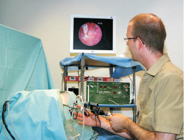
Surgical simulators present a safe and potentially effective method for surgical training, and can also be used in robot-assisted surgery for pre- and intra-operative planning. Accurate modeling of the interaction between surgical instruments and organs has been recognized as a key requirement in the development of high-fidelity surgical simulators. Researchers have attempted to model tool-tissue interactions in a wide variety of ways, which can be broadly classified as (1) linear elasticity-based, (2) nonlinear (hyperelastic) elasticity-based finite element (FE) methods, and (3) other techniques that not based on FE methods or continuum mechanics. Realistic modeling of organ deformation requires populating the model with real tissue data (which are difficult to acquire in vivo) and simulating organ response in real time (which is computationally expensive). Further, it is challenging to account for connective tissue supporting the organ, friction, and topological changes resulting from tool-tissue interactions during invasive surgical procedures. Overcoming such obstacles will not only help us to model tool-tissue interactions in real time, but also enable realistic force feedback to the user during surgical simulation. This review paper classifies the existing research on tool-tissue interactions for surgical simulators specifically based on the modeling techniques employed and the kind of surgical operation being simulated, in order to inform and motivate future research on improved tool-tissue interaction models.


Figures

Figure 1
Figure 1
Hysteroscopy training simulation environment coupled with a haptic device (Harders et al., 2006). Reprinted from Studies in Health Technology and Informatics, Vol. 119, Harders et al. Highly-realistic, immersive training environment for hysteroscopy, pp. 176–181, ©(2006), with permission from IOS Press.
Figure 2
Figure 2
Tissue fiber orientation of the heart on the inside surface, (a) and (b), and outside surface, (c) and (d), constructed using diffusion tensor imaging (Zhukov & Barr, 2003). Images are printed with permission from ©IEEE 2003.
Figure 3
Figure 3
Examples of characteristic properties of viscoelastic materials (a) creep and creep recovery - for a constant applied shear stress σ0 results in an increase in shear strain (b) stress relaxation - for a constant applied shear strain ε0 results in a decrease in shear stress until it reaches a steady state value.
Figure 4
Figure 4
Standard viscoelastic models commonly used to represent soft tissues (a) Maxwell (b) Kelvin-Voigt (or Voigt) (c) Zener standard linear solid (or Kelvin) (Fung, 1993).
Figure 5
Figure 5
Two-dimensional ABAQUS simulation results for soft tissue deformation of the human kidney that incorporates a hyperelastic constitutive model (Mooney-Rivlin model: C10 = 682.31 Pa and C01 = 700.02 Pa (Kim & Srinivasan, 2005)) and the left side boundary nodes are fixed, while loads are applied at the bottom and right edge nodes (a) undeformed mesh (b) contour plot of displacements (c) undeformed mesh is black, while deformed mesh is red.
Figure 6
Figure 6
(a) Tools tested and measuring tool-material interaction forces during (b) and (c) deformation of rubber (d) deformation of bovine liver (Mahvash et al., 2002). Images printed with permission from publisher (EuroHaptics 2002).
Figure 7
Figure 7
Indentation test on the “Truth Cube” embedded with fiducials (a) experimental test setup (b) CT of center vertical slice under 22% strain (c) FE model under 22% strain (Kerdok et al., 2003). Images printed with permission from Copyright ©2003 Elsevier B.V.
Figure 8
Figure 8
Formulation and results of the endoscopic simulator: (a) FE model of the human uterus containing 2000 elements. (b) Tool-tissue interaction model used in the surgical simulator (Székely et al., 2000). Images printed with permission from MIT Press Journals ©2000 by the Massachusetts Institute of Technology.
Figure 9
Figure 9
Needle insertion and simulation modeling: (a) Probing for estimation of material properties of phantom tissue. (b) 17 gauge epidural needle inserted into phantom tissue while motion of markers and insertion forces are recorded. (c) FE simulation of needle insertion with small target embedded within elastic tissue (DiMaio & Salcudean, 2003a). Images printed with permission from ©IEEE 2003.
Figure 10
Figure 10
Results from work presented in Picinbono et al. (2003): (a) Comparison between linear (wireframe) versus nonlinear (solid) elasticity-based models for same force applied to right lobe of the liver; the linear model undergoes large unrealistic deformation. (b) Simulating hepatic resection using a nonlinear anisotropic model. Images printed with permission from Copyright ©2003 Elsevier Science (USA).
Figure 11
Figure 11
Devices used to measure tissue properties in vivo: (a) Tissue aspiration technique (Vuskovic et al., 2000). Image printed with permission from ©IEEE 2000. (b) TeMPeST 1-D with 12 mm surgical port (Ottensmeyer, 2002). Image printed with permission from Wiley-Blackwell Publishing Ltd.
Figure 12
Figure 12
Modeling the information flow in simulator development and application. Each stage acts as a “filter” in which information about force-motion relationships are lost or transformed. Images are obtained from Roberts et al. (2007), Eidgenössische Technische Hochschule, Sacred Heart Medical Center (2004), SensAble Technologies Inc. (1990), and Institut National de Recherche en Informatique et en Automatique. Image corresponding to Roberts et al. (2007) printed with permission from Copyright ©2007 Elsevier Ltd.

References

    1. ADINA R & D Inc. (1986). 71 Elton Avenue, Watertown, MA 02472 USA. (http://www.adina.com/)

    1. Alterovitz R, Goldberg K, Pouliot J, Taschereau R, Hsu CI. Needle insertion and radioactive seed implantation in human tissues: simulation and sensitivity analysis. Proc. IEEE Int’l. Conf. on Robotics and Automation; Taipei, Taiwan. 2003. pp. 1793–1799.
    1. ANSYS Inc. (1970). Southpointe, 275 Technology Drive, Canonsburg, PA 15317 USA. (http://www.ansys.com/)

    1. Arruda EM, Boyce MC. Three-dimensional constitutive model for the large stretch behavior of rubber elastic materials. Journal of the Mechanics and Physics of Solids. 1993;41(2):389–412.
    1. Balaniuk R, Salisbury K. Dynamic simulation of deformable objects using the long elements method. Proc. 10th Symposium on Haptic Interfaces for Virtual Environments and Teleoperator Systems; Orlando, USA. 2002. pp. 58–65.

LinkOut - more resources