. 2010 May;119(5):617-30.
doi: 10.1007/s00401-010-0644-7. Epub 2010 Feb 2.

Canine distemper virus persistence in demyelinating encephalitis by swift intracellular cell-to-cell spread in astrocytes is controlled by the viral attachment protein

Affiliations

Canine distemper virus persistence in demyelinating encephalitis by swift intracellular cell-to-cell spread in astrocytes is controlled by the viral attachment protein

Gaby Wyss-Fluehmann et al. Acta Neuropathol. 2010 May.

Abstract

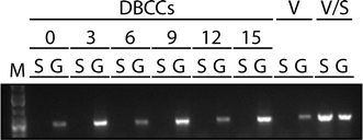
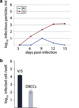
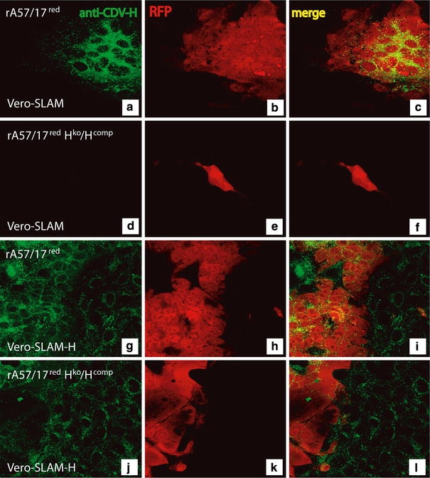
The mechanism of viral persistence, the driving force behind the chronic progression of inflammatory demyelination in canine distemper virus (CDV) infection, is associated with non-cytolytic viral cell-to-cell spread. Here, we studied the molecular mechanisms of viral spread of a recombinant fluorescent protein-expressing virulent CDV in primary canine astrocyte cultures. Time-lapse video microscopy documented that CDV spread was very efficient using cell processes contacting remote target cells. Strikingly, CDV transmission to remote cells could occur in less than 6 h, suggesting that a complete viral cycle with production of extracellular free particles was not essential in enabling CDV to spread in glial cells. Titration experiments and electron microscopy confirmed a very low CDV particle production despite higher titers of membrane-associated viruses. Interestingly, confocal laser microscopy and lentivirus transduction indicated expression and functionality of the viral fusion machinery, consisting of the viral fusion (F) and attachment (H) glycoproteins, at the cell surface. Importantly, using a single-cycle infectious recombinant H-knockout, H-complemented virus, we demonstrated that H, and thus potentially the viral fusion complex, was necessary to enable CDV spread. Furthermore, since we could not detect CD150/SLAM expression in brain cells, the presence of a yet non-identified glial receptor for CDV was suggested. Altogether, our findings indicate that persistence in CDV infection results from intracellular cell-to-cell transmission requiring the CDV-H protein. Viral transfer, happening selectively at the tip of astrocytic processes, may help the virus to cover long distances in the astroglial network, "outrunning" the host's immune response in demyelinating plaques, thus continuously eliciting new lesions.


Figures

Fig. 1
Fig. 1
Spread of rA75/17red in primary dog brain cell cultures (DBCCs). ad The recombinant A75/17-CDV, expressing an additional red fluorescent protein (rA75/17red), efficiently spreads in DBCCs. A growing infected focus is shown at 3, 6, 9 and 12 dpi. Asterisk depicts the same coordination point throughout the time of infection. Red fluorescence emission outlines the whole infected cells and their processes. Infected cells are widely spaced but seem always in contact with each other by way of their processes. Confocal laser microscopy ×20. e rA75/17red-infected DBCCs are in contact through thin cell processes (white arrowheads), as observed by the red fluorescence emission. Confocal laser microscopy ×200. f Merged images of CDV-induced red fluorescence emission and corresponding DIC image demonstrating that the DBCCs are confluent with spread of CDV from infected to target cells bypassing numerous uninfected cells. Confocal laser microscopy ×200. g Merged images of CDV nucleoproteins (N) labeled with a primary anti-N antibody, followed by a secondary alexa fluor 488-conjugated antibody (green), with red fluorescence emission in rA75/17red-infected cells (red) shows co-localization of both labels (yellow) excluding passive diffusion of RFP. Confocal laser microscopy ×200. h Astrocytes are the main target of rA75/17red in DBCCs. Merged images of CDV-infected cells by direct emission of red fluorescence (red) and astrocytes stained with an anti-GFAP antibody, followed by a secondary alexa fluor 555-conjugated antibody. i The CDV nucleocapsids (green) are located both in the cytosol and processes of infected astrocytes (red). Merged images of CDV nucleoprotein (N) stained with an anti-N antibody, followed by a secondary alexa fluor 488-conjugated antibody and astrocytes stained with an anti-GFAP antibody, followed by a secondary alexa fluor 555-conjugated antibody. Confocal laser microscopy ×200. j 3D reconstruction of 30 Z stacking images taken by confocal laser microscopy illustrating the presence of CDV nucleocapsids [anti-N antibody staining, followed by alexa fluor 488-conjugated antibody (green)] inside thin long astrocytic processes [anti-GFAP antibody staining, followed by alexa fluor 555-conjugated antibody (red)]. Confocal laser microscopy ×1,000
Fig. 2
Fig. 2
Video microscopy of DBCCs infected with rA75/17red. Consecutive images of an infected focus taken at 1 h intervals (or 18 h; two first images). Red arrowhead depicts an infected cell at the beginning of the sequence (72 h post-infection). From 90 hpi, the infection is spreading to other cells by contact of cell processes in various directions. Additional cells become fluorescent within intervals of 1 h. The yellow arrowhead depicts one of these cells (91 hpi). A process of the latter becomes fluorescent at 95 hpi. One hour later (96 hpi), the latter is in contact with a process of an additional cell (depicted by blue arrowhead), now becoming fluorescent. The last image of the series (104 hpi) illustrates an overlay of the red fluorescent protein with phase contrast showing a confluent culture, thereby demonstrating that the infection spreads selectively bypassing numerous cells which remain uninfected (×40)
Fig. 3
Fig. 3
The universal Morbillivirus CD150/SLAM receptor is not expressed in primary canine brain cells. DBCCs were infected (or left uninfected) with rA75/17red at an MOI of 0.01 (as titrated in Vero-SLAM cells). Total RNA was then extracted and subjected to RT-PCRs for assessing SLAM and the housekeeping gene GAPDH expressions at the indicated days post-infection. In addition, total RNA was extracted from Vero and Vero-SLAM cells to control for any defect in the RT-PCR system. S SLAM gene, G GAPDH gene, V Vero cells, V/S Vero-SLAM cells
Fig. 4
Fig. 4
rA75/17red is defective in free particles formation in primary canine brain cells. a DBCCs were infected with rA75/17red at an MOI of 0.01 (as titrated in Vero-SLAM cells). At the indicated days post-infection, cell-free (SN) and cell-associated viruses (CA) were harvested and titrated. Virus titers were determined 3 dpi by counting the number of virus-infected cells (RFP-expressing cells). The cells were overlaid 2 h post-infection with medium containing 1% of soft agar. Values represent the mean of three independent experiments. b Virus entry efficiency. Vero-SLAM cells (V/S) and DBCCs were infected with rA75/17red at an MOI of 0.01 (titrated in Vero-SLAM cells). One day post-infection, the number of infected Vero-SLAM cells and DBCCs per well were counted and are represented in the graph
Fig. 5
Fig. 5
Ultrastructural images of DBCCs infected with A75/17-CDV. a Large and small cell adjacent processes containing cross and longitudinally sectioned tubular profiles of CDV nucleocapsids (arrowheads). AF bundles of astrocytic fibrils (×11,000). b Two adjacent astroglial cell processes. In the cytosol of the left process accumulation of “fuzzy” nucleocapsids (open arrowhead) close to cell membrane with focal blurring of the adjacent cell membranes between the two cells (×15,000). c Two adjacent cell processes. The upper process contains an electron-dense nucleocapsid aggregate. Discontinuous plasmalemmal densities, “spiking” (filled arrowheads), distinction between adjacent cell membranes becomes less clear in this area. Arrow depicts nearby gap junction (×11,000). d Cell process between two cell bodies containing electron-dense nucleocapsid aggregates with tubular structures embedded in a granular matrix (arrowheads) attached to the cell membrane (×11,000). e Several closely spaced cell processes, containing nucleocapsid aggregates (open arrowheads). Several plasmalemmal densities (grey arrowheads), one of which exhibits “spiking” morphology (double arrowhead) (×11,000). f Low power composite of four overlapping EM photographs showing an astrocyte with nucleus (A1) and parts of the perikaryon of two further astrocytes (A2, A3) all of which contain numerous nucleocapsid aggregates (a few depicted by white arrowheads) (×3,000). In addition, the image shows several large and small cell processes equally containing nucleocapsid aggregates and one large viral inclusion (double white arrowhead). Small arrows depict a cell process connecting A2 and A3 with continuity between the cytoplasm of both perikarya
Fig. 6
Fig. 6
The CDV fusogenic complexes are potentially functional in DBCCs. a, b Both CDV surface glycoproteins are properly expressed in glial cells. DBCCs were infected with rA75/17red with an MOI of 0.01. Merged images of both glycoproteins F and H stained with anti-F and anti-H antibodies, respectively, followed by secondary alexa fluor 488-conjugated antibody (green) and astrocytes labeled using an anti-GFAP antibody, followed by a secondary alexa fluor 555-conjugated antibody (red). Confocal laser microscopy ×200. c, d F/H complexes are cell surface targeted in potentially functional. Prior to infection of DBCCs with rA75/17red (MOI of 0.01), cells were transduced with a lentivirus vector expressing SLAM (pRRL-SLAM) or with a control empty plasmid (pRRL). Syncytia formation was readily observed in CDV-infected cells. No syncytia are demonstrated in the control culture
Fig. 7
Fig. 7
Characterization of the recombinant Hko rA75/17red virus. Vero-SLAM (af) and Vero-SLAM-H cells (gi) were infected with rA75/17red and rA75/17red/Hko/Hcomp. One day post-infection, both cells were fixed, permeabilized and stained using the anti-H MAb 1C42H11 (VMRD) to detect the CDV hemagglutinin protein. This was followed by addition of an alexa fluor 488-conjugated secondary antibody. In addition, auto-fluorescence of the RFP is also shown, which allowed us for a direct illumination of infected cells. Merged images are also represented (c, f, i and l). Fluorescent images were taken using a confocal scanning laser microscope (Olympus) (×200)
Fig. 8
Fig. 8
Role of the receptor-binding H protein in viral spread. ad Infection of DBCCs with a hemagglutinin (H) knockout, H trans-complemented recombinant A75/17 virus (rA75/17red/Hko/Hcomp). The latter recombinant virus required initial trans-complementation with the wild-type hemagglutinin (Hwt) protein in Vero-SLAM cells stably expressing Hwt (Vero-SLAM-H cells). The four images taken at 3, 6, 9 and 12 dpi of the same infected focus illustrated no further spread from the initially infected cells. Infected cells were identified by capturing red fluorescence emission using a confocal laser microscope. e, f DBCCs infected with rA75/17red and treated with an antibody against H (MAb 1347 [26]) exhibited markedly fewer infected cells as compared to cultures treated with a control antibody (MAb anti-HA). Infected cells were identified by capturing red fluorescence emission using a confocal laser microscope

Similar articles

Cited by

References

    1. Bollo E, Zurbriggen A, Vandevelde M, Fankhauser R. Canine distemper virus clearance in chronic inflammatory demyelination. Acta Neuropathol (Berl) 1986;72:69–73. doi: 10.1007/BF00687949. - DOI - PubMed
    1. Brettschneider J, Tumani H, Kiechle U, Muche R, Richards G, Lehmensiek V, Ludolph AC, Otto M. IgG antibodies against measles, rubella, and varicella zoster virus predict conversion to multiple sclerosis in clinically isolated syndrome. PLoS One. 2009;4:e7638. doi: 10.1371/journal.pone.0007638. - DOI - PMC - PubMed
    1. Brown HR, Goller N, Thormar H, Norrby E. Fuzzy material surrounding measles virus nucleocapsids identified as matrix protein. Brief report. Arch Virol. 1987;94:163–168. doi: 10.1007/BF01313735. - DOI - PubMed
    1. Buchholz UJ, Finke S, Conzelmann KK. Generation of bovine respiratory syncytial virus (BRSV) from cDNA: BRSV NS2 is not essential for virus replication in tissue culture, and the human RSV leader region acts as a functional BRSV genome promoter. J Virol. 1999;73:251–259. - PMC - PubMed
    1. Calain P, Roux L. The rule of six, a basic feature for efficient replication of Sendai virus defective interfering RNA. J Virol. 1993;67:4822–4830. - PMC - PubMed

Publication types

MeSH terms

Substances