

Differential expression of proteins in fetal brains of alcohol-treated prenatally C57BL/6 mice: a proteomic investigation
- PMID: 20119957
- PMCID: PMC3431165
- DOI: 10.1002/elps.200900385
Differential expression of proteins in fetal brains of alcohol-treated prenatally C57BL/6 mice: a proteomic investigation
Abstract
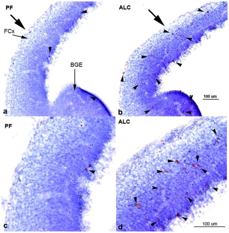
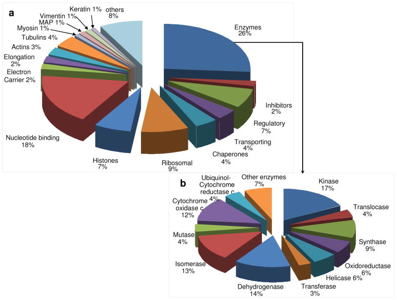
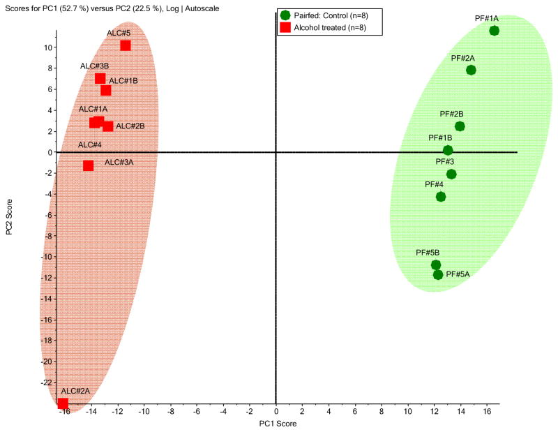
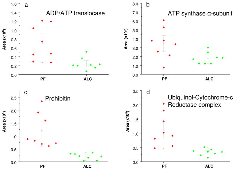
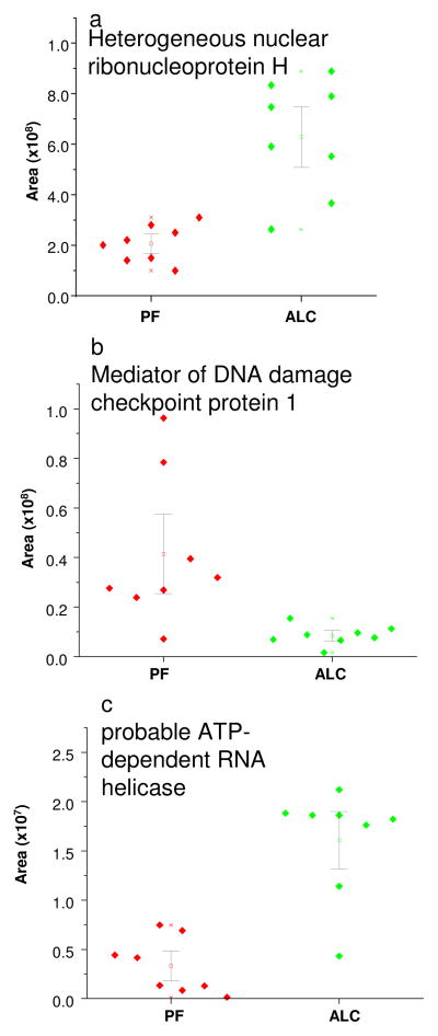
Alcohol is known to impede the growth of the central nervous system and to induce neurodegeneration through cellular apoptosis. We have previously shown that moderate prenatal alcohol exposure results in brain defects at different stages of development. In this study, we further characterize the proteomic architecture underlying ethanol teratogenesis during early fetal brain development using chromatography in conjunction with a LC-MS/MS system. Pregnant C57BL/6 mice were exposed from embryonic day 7 (E7) to E13 with either a 25% ethanol derived calorie or pair-fed liquid diets. At E13, fetal brains were collected from five dams for each group. Individual brains were homogenized and the extracted proteins were then tryptically digested and analyzed by LC-MS/MS. Label-free quantitative proteomic analyses were performed on proteomes extracted from fetal brains of both alcohol-treated (ALC) and pair-fed groups. These analyses demonstrated that prenatal alcohol exposure induced significant downregulation (p<0.001) of the expression of mitochondrial enzymes including ADP/ATP translocase 1, ATP synthase subunit alpha and ubiquinol-cytochrome-c reductases. In addition, mitochondrial carrier homolog 1, which plays a role in apoptosis, was significantly downregulated (p<0.001) in the ALC group. Moreover, among the cytosolic proteins that were significantly downregulated (p<0.001) are Bcl-2, 14-3-3 protein and calmodulin. Significant downregulation (p<0.001) of proteins that are critical for fetal brain development was observed such as prohibitin and neuronal migration protein doublecortin. These findings provide information about possible mechanisms underlying the effects of prenatal alcohol exposure during early embryonic stage.
Figures
Similar articles
-
Alteration of selective neurotransmitters in fetal brains of prenatally alcohol-treated C57BL/6 mice: quantitative analysis using liquid chromatography/tandem mass spectrometry.Int J Dev Neurosci. 2010 May;28(3):263-9. doi: 10.1016/j.ijdevneu.2010.01.004. Epub 2010 Feb 1. Int J Dev Neurosci. 2010. PMID: 20123123 Free PMC article.
-
Neuroprotective peptide ADNF-9 in fetal brain of C57BL/6 mice exposed prenatally to alcohol.J Biomed Sci. 2011 Oct 21;18(1):77. doi: 10.1186/1423-0127-18-77. J Biomed Sci. 2011. PMID: 22017746 Free PMC article.
-
Moderate alcohol exposure compromises neural tube midline development in prenatal brain.Brain Res Dev Brain Res. 2003 Aug 12;144(1):43-55. doi: 10.1016/s0165-3806(03)00158-5. Brain Res Dev Brain Res. 2003. PMID: 12888216
-
Intrauterine environment-genome interaction and children's development (1): Ethanol: a teratogen in developing brain.J Toxicol Sci. 2009;34 Suppl 2:SP273-8. doi: 10.2131/jts.34.sp273. J Toxicol Sci. 2009. PMID: 19571480 Review.
-
Fetal alcohol spectrum disorders and fetal alcohol syndrome: the state of the art and new diagnostic tools.Early Hum Dev. 2013 Jun;89 Suppl 1:S40-3. doi: 10.1016/S0378-3782(13)70013-6. Early Hum Dev. 2013. PMID: 23809349 Review.
Cited by
-
Fetal regional brain protein signature in FASD rat model.Reprod Toxicol. 2018 Mar;76:84-92. doi: 10.1016/j.reprotox.2018.01.004. Epub 2018 Feb 1. Reprod Toxicol. 2018. PMID: 29408587 Free PMC article.
-
Activity-dependent neuroprotective protein-derived peptide, NAP, preventing alcohol-induced apoptosis in fetal brain of C57BL/6 mouse.Neuroscience. 2009 Feb 18;158(4):1426-35. doi: 10.1016/j.neuroscience.2008.11.021. Epub 2008 Nov 21. Neuroscience. 2009. PMID: 19073235 Free PMC article.
-
Prohibitin reduces mitochondrial free radical production and protects brain cells from different injury modalities.J Neurosci. 2012 Jan 11;32(2):583-92. doi: 10.1523/JNEUROSCI.2849-11.2012. J Neurosci. 2012. PMID: 22238093 Free PMC article.
-
Alcohol-induced alterations in maternal uterine endothelial proteome: a quantitative iTRAQ mass spectrometric approach.Reprod Toxicol. 2012 Dec;34(4):538-44. doi: 10.1016/j.reprotox.2012.08.008. Epub 2012 Sep 5. Reprod Toxicol. 2012. PMID: 22960358 Free PMC article.
-
Experimental methods for testing the effects of neurotrophic peptide, ADNF-9, against alcohol-induced apoptosis during pregnancy in c57bl/6 mice.J Vis Exp. 2013 Apr 24;(74):e50092. doi: 10.3791/50092. J Vis Exp. 2013. PMID: 23644584 Free PMC article.
References
-
- Bauer-Moffett C, Altman J. Brain Res. 1977;119:249–268. - PubMed
-
- Kornguth SE, Rutledge JJ, Sunderland E, Siegel F, et al. Brain Res. 1979;177:347–360. - PubMed
-
- Samson HH, Diaz J. Alcohol Clin Exp Res. 1981;5:563–569. - PubMed
-
- Pierce DR, West JR. Alcohol. 1986;3:185–191. - PubMed
-
- Bonthius DJ, Goodlett CR, West JR. Alcohol. 1988;5:209–214. - PubMed
Publication types
MeSH terms
Substances
Grants and funding
LinkOut - more resources
Full Text Sources
Medical
