

doi: 10.1021/jm9012938.
Evolving carbapenemases: can medicinal chemists advance one step ahead of the coming storm?
Affiliations
- PMID: 20121112
- PMCID: PMC2855403
- DOI: 10.1021/jm9012938
Item in Clipboard
Evolving carbapenemases: can medicinal chemists advance one step ahead of the coming storm?
J Med Chem.
.
No abstract available
Figures
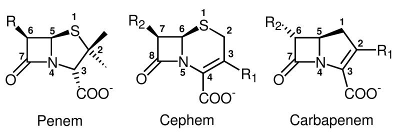
Chemical structures of the bicyclic cores of different classes of β-lactam antibiotics. The penem core is found in penicillins and consists of a β-lactam ring fused with a tetrahydrodrothiazole ring. The cephem core is found in cephalosporins and consists of a β-lactam ring fused with a dihydrothiazine ring. The carbapenem core consists of a β-lactam ring fused with a dihydropyrrole ring. Heavy atoms of the bicyclic systems are numbered according to common use rather than according to the IUPAC nomenclature to facilitate comparisons between the different antibiotics. Note that the numbering of the R groups is arbitrary; here we started labeling R groups from the ones attached to the core atoms with the lowest number.
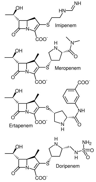
Chemical structures of four commonly prescribed carbapenems: imipenem ((5R,6S)-3-[2-(aminomethylideneamino)ethylsulfanyl]-6-(1-hydroxyethyl)-7-oxo-1-azabicyclo[3.2.0]hept-2-ene-2-carboxylic acid), meropenem (3-[5-(dimethylcarbamoyl)pyrrolidin-2-yl] sulfanyl-6- (1-hydroxyethyl)-4-methyl-7-oxo- 1-azabicyclo[3.2.0] hept-2-ene-2-carboxylic acid); ertapenem ((4R,5S,6S)-3-[(3S,5S)-5-[(3-carboxyphenyl)carbamoyl]pyrrolidin-3-yl]sulfanyl-6-(1-hydroxyethyl)-4-methyl-7-oxo-1-azabicyclo[3.2.0]hept-2-ene-2-carboxylic acid); and doripenem ((4R,5S,6S)-6-(1-hydroxyethyl)-4-methyl-7-oxo-3-[(3S,5S)-5-[(sulfamoylamino)methyl]pyrrolidin-3-yl]sulfanyl-1-azabicyclo[3.2.0]hept-2-ene-2-carboxylic acid).
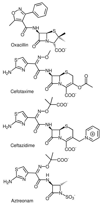
Chemical structures of selected non-carbapenem β-lactam antibiotics in clinical use: oxacillin ((2S,5R,6R)-3,3-dimethyl-6-[(5-methyl-3-phenyl-1,2-oxazole-4-carbonyl)amino]-7-oxo-4-thia-1-azabicyclo[3.2.0]heptane-2-carboxylic acid), a penicillin; cefotaxime ((6R,7R,Z)-3-(acetoxymethyl)-7-(2-(2-aminothiazol-4-yl)-2-(methoxyimino)acetamido)-8-oxo-5-thia-1-azabicyclo[4.2.0]oct-2-ene-2-carboxylic acid), a third generation cephalosporin; ceftazidime ((6R,7R,Z)-7-(2-(2-aminothiazol-4-yl)-2-(2-carboxypropan-2-yloxyimino)acetamido)-8-oxo-3-(pyridinium-1-ylmethyl)-5-thia-1-aza-bicyclo[4.2.0]oct-2-ene-2-carboxylate), another third generation cephalosporin; and aztreonam (2-(([(1Z)-1-(2-amino-1,3-thiazol-4-yl)-2-([(2S,3S)-2-methyl-4-oxo-1-sulfoazetidin-3-yl]amino)-2-oxoethylidene]amino)oxy)-2-methylpropanoic acid), a monocyclic β-lactam or monobactam.
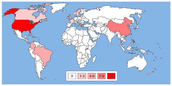
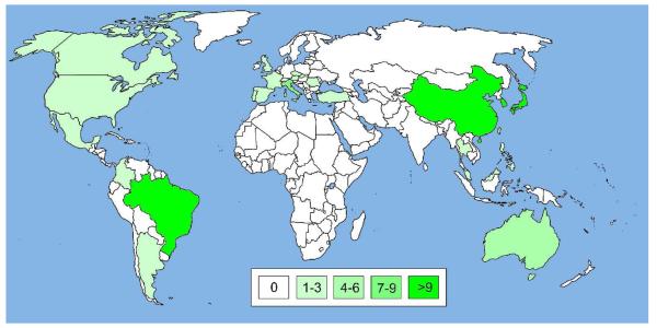
World map illustrating the global spread of KPC enzymes. A blank world map was obtained from http://upload.wikimedia.org/ and countries with KPC occurences were colored in different opacities of red (symbolizing SBLs) according to the number of publications found on PubMed at http://www.ncbi.nlm.nih.gov/ . Publications were retrieved using search strings such as “KPC-* United States” and titles and abstracts were checked for content. Only articles reporting occurences of KPCs were included, while review articles and reports restricted to computational and/or in vitro studies were excluded. Countries, for which ten or more publications with KPC reports were found, were colored in red with 100% opacity; those with fewer publications with lower opacities: 7-9 publications, 80%; 4-6 publications, 60%; 1-3 publications, 40%; no publications, white (see color code in the Figure). For more details see Supporting Information S2-S3.
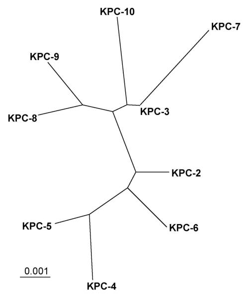
Radial phylogenetic tree of currently known KPC enzymes. Amino acid sequences of KPC enzymes including the leader sequence were retrieved from GenBank at http://www.ncbi.nlm.nih.gov/ and aligned using Clustal X Version 2.0.9 using default parameters. The phylogenetic tree was visualized using TreeView. The bar at the lower left corner gives a measure for amino acid sequence diversity. For instance, two enzymes differing by only one of 293 amino acid residues share 99.66% sequence identity and differ by 0.34% (0.0034). The KPC-9 sequence was missing five and four residues at the N- and C-termini, respectively. Since these residues are 100% conserved in the other enzymes, we added the missing residues accordingly. For more details see Supporting Information S2-S3.
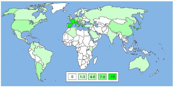
World map illustrating the global spread of IMP enzymes. The map was prepared as described for Figure 4 except that IMP-specific search strings were used to retrieve articles and that countries with IMP occurence were colored in green, symbolizing MBLs. In some cases, where no published articles were available, GenBank entries were taken into account, e.g., for Thailand. For more details see Supporting Information S3-S5.
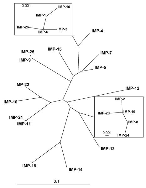
Radial phylogenetic tree of currently known IMP enzymes, generated as described for Figure 5. Two clusters of closely related enzymes, the “IMP-1 cluster” and “IMP-2 cluster” are shown as insets (note the different scale of the sequence diversity measures). For more details see Supporting Information S3-S5.
World map illustrating the global spread of VIM enzymes. The map was prepared as described for Figure 6 except that VIM-specific search strings were used to retrieve articles. For more details see Supporting Information S6-S8.
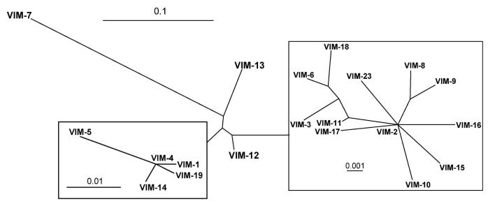
Radial phylogenetic tree of currently known VIM enzymes, generated as described for Figure 5. For more details see Supporting Information S6-S8.
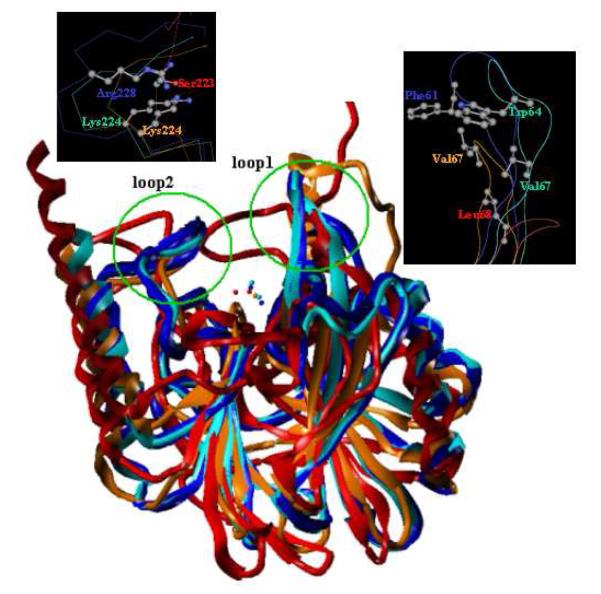
Superimposition of crystal structures of MBLs from three subclasses. Selected enzymes include VIM-2 (subclass B1, blue, PDB code 2YZ3), IMP-1 (subclass B1, cyan, PDB code, 1DD6), CphA (subclass B2, orange, PDB code 2QDS), and L1 (subclass B3, red, PDB code 2AIO). Two flexible loops are highlighted by green circles. Important residues in the two loops are depicted and colored by atom type (carbon, grey; oxygen, red; nitrogen, blue). Clearly, residues in loop1 are hydrophobic, while those in loop2 possess mostly positively charged side chains.
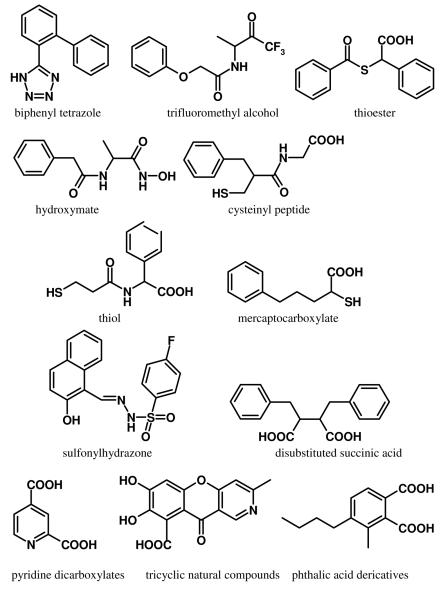
Chemical structures of selected MBL inhibitors.
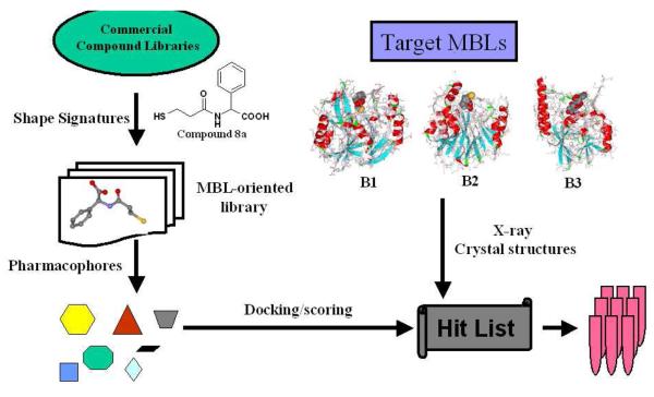
Proposed VS scheme for the discovery of MBL inhibitors with broad-spectrum activities against B1, B2, and B3 subclasses.
Similar articles
-
Crystal structures of KPC-2 β-lactamase in complex with 3-nitrophenyl boronic acid and the penam sulfone PSR-3-226.Antimicrob Agents Chemother. 2012 May;56(5):2713-8. doi: 10.1128/AAC.06099-11. Epub 2012 Feb 13. Antimicrob Agents Chemother. 2012. PMID: 22330909 Free PMC article.
-
Local interactions with the Glu166 base and the conformation of an active site loop play key roles in carbapenem hydrolysis by the KPC-2 β-lactamase.J Biol Chem. 2021 Jan-Jun;296:100799. doi: 10.1016/j.jbc.2021.100799. Epub 2021 May 20. J Biol Chem. 2021. PMID: 34022225 Free PMC article.
-
Klebsiella pneumoniae Carbapenemase-2 (KPC-2), Substitutions at Ambler Position Asp179, and Resistance to Ceftazidime-Avibactam: Unique Antibiotic-Resistant Phenotypes Emerge from β-Lactamase Protein Engineering.mBio. 2017 Oct 31;8(5):e00528-17. doi: 10.1128/mBio.00528-17. mBio. 2017. PMID: 29089425 Free PMC article.
-
Ten Years with New Delhi Metallo-β-lactamase-1 (NDM-1): From Structural Insights to Inhibitor Design.ACS Infect Dis. 2019 Jan 11;5(1):9-34. doi: 10.1021/acsinfecdis.8b00247. Epub 2018 Nov 28. ACS Infect Dis. 2019. PMID: 30421910 Review.
-
Decoding the Structural Basis For Carbapenem Hydrolysis By Class A β-lactamases: Fishing For A Pharmacophore.Curr Drug Targets. 2016;17(9):983-1005. doi: 10.2174/1389450116666151001104448. Curr Drug Targets. 2016. PMID: 26424401 Review.
Cited by
-
Protein variants form a system of networks: microdiversity of IMP metallo-beta-lactamases.PLoS One. 2014 Jul 11;9(7):e101813. doi: 10.1371/journal.pone.0101813. eCollection 2014. PLoS One. 2014. PMID: 25013948 Free PMC article.
-
Carbamylmethyl Mercaptoacetate Thioether: A Novel Scaffold for the Development of L1 Metallo-β-lactamase Inhibitors.ACS Med Chem Lett. 2017 Apr 24;8(5):527-532. doi: 10.1021/acsmedchemlett.7b00058. eCollection 2017 May 11. ACS Med Chem Lett. 2017. PMID: 28523105 Free PMC article.
-
On drug discovery against infectious diseases and academic medicinal chemistry contributions.Beilstein J Org Chem. 2022 Sep 29;18:1355-1378. doi: 10.3762/bjoc.18.141. eCollection 2022. Beilstein J Org Chem. 2022. PMID: 36247982 Free PMC article.
-
Elucidating the Role of Residue 67 in IMP-Type Metallo-β-Lactamase Evolution.Antimicrob Agents Chemother. 2015 Dec;59(12):7299-307. doi: 10.1128/AAC.01651-15. Epub 2015 Sep 14. Antimicrob Agents Chemother. 2015. PMID: 26369960 Free PMC article.
-
Assay platform for clinically relevant metallo-β-lactamases.J Med Chem. 2013 Sep 12;56(17):6945-53. doi: 10.1021/jm400769b. Epub 2013 Aug 16. J Med Chem. 2013. PMID: 23898798 Free PMC article.
References
-
- Rossolini GM, Mantengoli E. Treatment and control of severe infections caused by multiresistant Pseudomonas aeruginosan. Clin. Microbiol. Infect. 2005;11(Suppl 4):17–32. - PubMed
-
- Karageorgopoulos DE, Falagas ME. Current control and treatment of multidrug-resistant Acinetobacter baumannii infections. Lancet Infect. Dis. 2008;8:751–762. - PubMed
-
- Ramphal R, Ambrose PG. Extended-spectrum β-lactamases and clinical outcomes: current data. Clin. Infect. Dis. 2006;42(Suppl 4):S164–172. - PubMed
-
- Isturiz R. Global resistance trends and the potential impact on empirical therapy. Int. J. Antimicrob. Agents. 2008;32(Suppl 4):S201–206. - PubMed
-
- Barza M. Imipenem: first of a new class of β-lactam antibiotics. Ann. Intern. Med. 1985;103:552–560. - PubMed
Publication types
MeSH terms
Substances
Grants and funding
LinkOut - more resources
Full Text Sources
Other Literature Sources
Medical
