Combined blood/tissue analysis for cancer biomarker discovery: application to renal cell carcinoma
- PMID: 20121140
- PMCID: PMC3251958
- DOI: 10.1021/ac902204k
Combined blood/tissue analysis for cancer biomarker discovery: application to renal cell carcinoma
Abstract
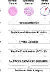
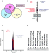
A method that relies on subtractive tissue-directed shot-gun proteomics to identify tumor proteins in the blood of a patient newly diagnosed with cancer is described. To avoid analytical and statistical biases caused by physiologic variability of protein expression in the human population, this method was applied on clinical specimens obtained from a single patient diagnosed with nonmetastatic renal cell carcinoma (RCC). The proteomes extracted from tumor, normal adjacent tissue and preoperative plasma were analyzed using 2D-liquid chromatography-mass spectrometry (LC-MS). The lists of identified proteins were filtered to discover proteins that (i) were found in the tumor but not normal tissue, (ii) were identified in matching plasma, and (iii) whose spectral count was higher in tumor tissue than plasma. These filtering criteria resulted in identification of eight tumor proteins in the blood. Subsequent Western-blot analysis confirmed the presence of cadherin-5, cadherin-11, DEAD-box protein-23, and pyruvate kinase in the blood of the patient in the study as well as in the blood of four other patients diagnosed with RCC. These results demonstrate the utility of a combined blood/tissue analysis strategy that permits the detection of tumor proteins in the blood of a patient diagnosed with RCC.
Conflict of interest statement
COMPETING INTERESTS STATEMENT
The authors declare that they have no competing financial interests.
Figures


Similar articles
-
Mass spectrometry in cancer biomarker research: a case for immunodepletion of abundant blood-derived proteins from clinical tissue specimens.Biomark Med. 2014;8(2):269-86. doi: 10.2217/bmm.13.101. Biomark Med. 2014. PMID: 24521024 Free PMC article.
-
Use of quantitative shotgun proteomics to identify fibronectin 1 as a potential plasma biomarker for clear cell carcinoma of the kidney.Cancer Biomark. 2011-2012;10(3-4):175-83. doi: 10.3233/CBM-2012-0243. Cancer Biomark. 2011. PMID: 22674303
-
Proteomic identification of Reticulocalbin 1 as potential tumor marker in renal cell carcinoma.J Proteomics. 2013 Oct 8;91:385-92. doi: 10.1016/j.jprot.2013.07.018. Epub 2013 Jul 31. J Proteomics. 2013. PMID: 23916412
-
Role of leptin as a biomarker for early detection of renal cell carcinoma? No evidence from a systematic review and meta-analysis.Med Hypotheses. 2019 Aug;129:109239. doi: 10.1016/j.mehy.2019.109239. Epub 2019 May 20. Med Hypotheses. 2019. PMID: 31371068
-
Diagnostic role of kidney injury molecule-1 in renal cell carcinoma.Int Urol Nephrol. 2019 Nov;51(11):1893-1902. doi: 10.1007/s11255-019-02231-0. Epub 2019 Aug 5. Int Urol Nephrol. 2019. PMID: 31385177 Review.
Cited by
-
Biomarker Analysis of Formalin-Fixed Paraffin-Embedded Clinical Tissues Using Proteomics.Biomolecules. 2023 Jan 3;13(1):96. doi: 10.3390/biom13010096. Biomolecules. 2023. PMID: 36671481 Free PMC article. Review.
-
Proteomic profiling of human plasma and intervertebral disc tissue reveals matrisomal, but not plasma, biomarkers of disc degeneration.Arthritis Res Ther. 2025 Feb 10;27(1):28. doi: 10.1186/s13075-025-03489-9. Arthritis Res Ther. 2025. PMID: 39930483 Free PMC article.
-
Comparative proteomics of a model MCF10A-KRasG12V cell line reveals a distinct molecular signature of the KRasG12V cell surface.Oncotarget. 2016 Dec 27;7(52):86948-86971. doi: 10.18632/oncotarget.13566. Oncotarget. 2016. PMID: 27894102 Free PMC article.
-
Analysis and interpretation of transcriptomic data obtained from extended Warburg effect genes in patients with clear cell renal cell carcinoma.Oncoscience. 2015 Feb 17;2(2):151-86. doi: 10.18632/oncoscience.128. eCollection 2015. Oncoscience. 2015. PMID: 25859558 Free PMC article.
-
Discovery of mouse spleen signaling responses to anthrax using label-free quantitative phosphoproteomics via mass spectrometry.Mol Cell Proteomics. 2011 Mar;10(3):M110.000927. doi: 10.1074/mcp.M110.000927. Epub 2010 Dec 28. Mol Cell Proteomics. 2011. PMID: 21189417 Free PMC article.
References
Publication types
MeSH terms
Substances
Grants and funding
LinkOut - more resources
Full Text Sources
Medical

