. 2010 Sep 15;13(6):731-43.
doi: 10.1089/ars.2009.2968.

A model of redox kinetics implicates the thiol proteome in cellular hydrogen peroxide responses

Affiliations

A model of redox kinetics implicates the thiol proteome in cellular hydrogen peroxide responses

Nnenna J Adimora et al. Antioxid Redox Signal. .

Abstract

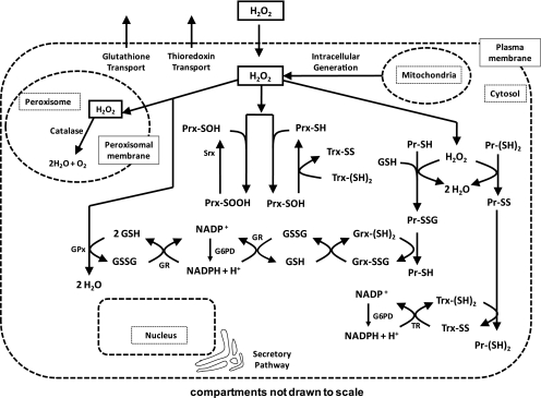
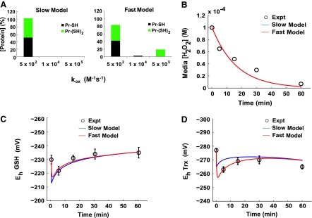
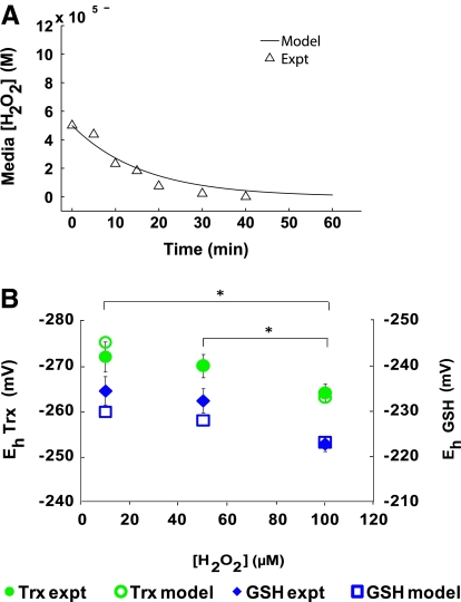
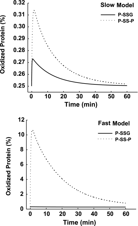
Hydrogen peroxide is appreciated as a cellular signaling molecule with second-messenger properties, yet the mechanisms by which the cell protects against intracellular H(2)O(2) accumulation are not fully understood. We introduce a network model of H(2)O(2) clearance that includes the pseudo-enzymatic oxidative turnover of protein thiols, the enzymatic actions of catalase, glutathione peroxidase, peroxiredoxin, and glutaredoxin, and the redox reactions of thioredoxin and glutathione. Simulations reproduced experimental observations of the rapid and transient oxidation of glutathione and the rapid, sustained oxidation of thioredoxin on exposure to extracellular H(2)O(2). The model correctly predicted early oxidation profiles for the glutathione and thioredoxin redox couples across a range of initial extracellular [H(2)O(2)] and highlights the importance of cytoplasmic membrane permeability to the cellular defense against exogenous sources of H(2)O(2). The protein oxidation profile predicted by the model suggests that approximately 10% of intracellular protein thiols react with hydrogen peroxide at substantial rates, with a majority of these proteins forming protein disulfides as opposed to protein S-glutathionylated adducts. A steady-state flux analysis predicted an unequal distribution of the intracellular anti-oxidative burden between thioredoxin-dependent and glutathione-dependent antioxidant pathways, with the former contributing the majority of the cellular antioxidant defense due to peroxiredoxins and protein disulfides.


Figures

FIG. 1.
FIG. 1.
Model of hydrogen peroxide (H2O2) elimination by Jurkat T cells. The modeled system was that of an individual cell with three compartments being considered: the extracellular medium, the intracellular cytosol, and the peroxisomes. H2O2 freely moved between compartments by permeating through the cytoplasmic and peroxisomal membranes. A constant intracellular H2O2 production rate was defined with a mitochondrial source. Within the intracellular cytosol, H2O2 was metabolized by a series of reactions (arrows), each with a given reaction-rate constant (Table 1). By four main pathways, H2O2 was metabolized by the cell. The first was controlled by catalase, and the second was controlled by glutathione peroxidase (GPx) enzymes working in conjunction with GSH, GR, and NADPH. The third was controlled by the peroxiredoxin enzymes (Prx) working in conjunction with Trx, TR, and NADPH. The fourth and final pathway described the nonenzymatic elimination of H2O2 through the oxidation of cysteine residues on intracellular proteins. This can occur at single Cys residues, resulting in sulfenic acids, which react with GSH to form glutathionylated intermediates. Alternatively, this can occur at dithiols resulting in the formation of an internal disulfide.
FIG. 2.
FIG. 2.
A model with rapid protein dithiol oxidation accurately describes hydrogen peroxide consumption. (A) Assignment of the distribution of protein thiol pools and rate constants for the slow and fast protein thiol oxidation models. (B) Experimental data (3) and model fitted results for the slow and fast protein thiol oxidation models showing the consumption of H2O2 by intact Jurkat T cells at a density of a 1 × 106 cells/ml after being exposed to a bolus addition of 100 μM extracellular H2O2. (C) Redox states of the glutathione redox couple as experimentally determined and model fitted for the slow and fast models. Experimental values represent the mean ± SEM of three separate experiments. (D) Redox states of the thioredoxin redox couple as experimentally determined and model fitted for the slow and fast protein thiol oxidation models. Experimental values represent the mean ± SEM of three separate experiments.
FIG. 3.
FIG. 3.
Model validation for varied initial extracellular peroxide concentrations. (A) Model-simulated and experimentally determined (13) consumption of H2O2 by intact Jurkat T cells (cell density, 1 × 106 cells/ml) after being exposed to a bolus addition of 50 μM extracellular H2O2. (B) Model-simulated and experimentally determined glutathione and thioredoxin redox potentials for varying initial extracellular [H2O2] (Jurkat T-cell density, 1 × 106 cells/ml). *Asterisks indicate statistically significant differences (p < 0.05) with respect to the two hydrogen peroxide concentrations being compared.
FIG. 4.
FIG. 4.
Model-predicted intracellular [H2O2]. Dynamic model-simulated extra- (top) and intracellular (bottom) peroxide concentration as a result of 100 μM H2O2. (Jurkat T-cell density = 1 × 106 cells/ml).
FIG. 5.
FIG. 5.
Model-predicted protein-oxidation profiles. Simulated percentages of S-glutathionylated proteins and protein disulfides that form as a result of oxidative stress (100 μM H2O2; Jurkat T-cell density = 1 × 106 cells/ml) for the slow (top) and fast (bottom) models.

Similar articles

Cited by

References

    1. Aebi H. Catalase in vitro. Methods Enzymol. 1984;105:121–126. - PubMed
    1. Akerboom TP. Bilzer M. Sies H. The relationship of biliary glutathione disulfide efflux and intracellular glutathione disulfide content in perfused rat liver. J Biol Chem. 1982;257:4248–4252. - PubMed
    1. Antunes F. Cadenas E. Estimation of H2O2 gradients across biomembranes. FEBS Lett. 2000;475:121–126. - PubMed
    1. Arner ES. Zhong L. Holmgren A. Preparation and assay of mammalian thioredoxin and thioredoxin reductase. Methods Enzymol. 1999;300:226–239. - PubMed
    1. Bienert GP. Schjoerring JK. Jahn TP. Membrane transport of hydrogen peroxide. Biochim Biophys Acta. 2006;1758:994–1003. - PubMed

Publication types

LinkOut - more resources