

Pulmonary cyclooxygenase-1 (COX-1) and COX-2 cellular expression and distribution after respiratory syncytial virus and parainfluenza virus infection
- PMID: 20121401
- PMCID: PMC2883516
- DOI: 10.1089/vim.2009.0042
Pulmonary cyclooxygenase-1 (COX-1) and COX-2 cellular expression and distribution after respiratory syncytial virus and parainfluenza virus infection
Abstract
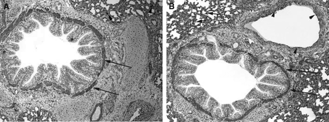
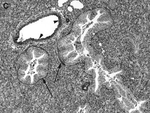
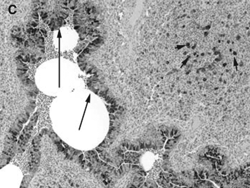
Prostaglandins (PGs) play an important role in pulmonary physiology and various pathophysiological processes following infection. The initial step in the biosynthesis of PGs is regulated by two distinct cyclooxygenase enzymes, cyclooxygenase-1 (COX-1) and COX-2. The goal of this study was to investigate the pulmonary cellular localization and distribution of COX-1 and COX-2 in a neonatal lamb model following respiratory syncytial virus (RSV) and parainfluenza virus 3 (PI3) infection, organisms that also cause significant respiratory disease in children. No significant differences were seen in pulmonary COX-1 expression at various microanatomical locations following RSV or PI3 infection compared to controls. In contrast, COX-2 was upregulated following RSV and PI3 infection. Strong expression was restricted to bronchial and bronchiolar epithelial cells and macrophages, while minimal expression was present in the same microanatomical locations in the uninfected lungs. Other microanatomical locations in both the controls and the infected lungs lacked expression. This work suggests that during RSV or PI3 infection: (1) COX-1 cellular expression is not altered, (2) COX-2 cellular expression is upregulated in airway bronchiolar and bronchial epithelial cells and macrophages, (3) respiratory epithelium along with macrophages are important microanatomical compartments regulating the host inflammatory response during viral infection, and (4) COX-2 may be a potential target for RSV and PI3 therapy.
Figures

Similar articles
-
Respiratory syncytial virus (RSV) infection induces cyclooxygenase 2: a potential target for RSV therapy.J Immunol. 2005 Apr 1;174(7):4356-64. doi: 10.4049/jimmunol.174.7.4356. J Immunol. 2005. PMID: 15778400
-
Kinetics of Respiratory Syncytial Virus (RSV) Memphis Strain 37 (M37) Infection in the Respiratory Tract of Newborn Lambs as an RSV Infection Model for Human Infants.PLoS One. 2015 Dec 7;10(12):e0143580. doi: 10.1371/journal.pone.0143580. eCollection 2015. PLoS One. 2015. PMID: 26641081 Free PMC article.
-
Respiratory syncytial virus infects the Bonnet monkey, Macaca radiata.Pediatr Dev Pathol. 1999 Jul-Aug;2(4):316-26. doi: 10.1007/s100249900129. Pediatr Dev Pathol. 1999. PMID: 10347274
-
Respiratory syncytial virus--viral biology and the host response.J Infect. 2002 Jul;45(1):18-24. doi: 10.1053/jinf.2002.1015. J Infect. 2002. PMID: 12217726 Review.
-
Respiratory syncytial virus persistence: evidence in the mouse model.Pediatr Infect Dis J. 2008 Oct;27(10 Suppl):S60-2. doi: 10.1097/INF.0b013e3181684d52. Pediatr Infect Dis J. 2008. PMID: 18820580 Review.
Cited by
-
Prostaglandin E2 induction during mouse adenovirus type 1 respiratory infection regulates inflammatory mediator generation but does not affect viral pathogenesis.PLoS One. 2013 Oct 16;8(10):e77628. doi: 10.1371/journal.pone.0077628. eCollection 2013. PLoS One. 2013. PMID: 24147040 Free PMC article.
-
Highly pathogenic porcine reproductive and respiratory syndrome virus induces prostaglandin E2 production through cyclooxygenase 1, which is dependent on the ERK1/2-p-C/EBP-β pathway.J Virol. 2014 Mar;88(5):2810-20. doi: 10.1128/JVI.03205-13. Epub 2013 Dec 18. J Virol. 2014. PMID: 24352469 Free PMC article.
-
Cyclooxygenase-2 inhibitor blocks the production of West Nile virus-induced neuroinflammatory markers in astrocytes.J Gen Virol. 2011 Mar;92(Pt 3):507-15. doi: 10.1099/vir.0.026716-0. Epub 2010 Nov 24. J Gen Virol. 2011. PMID: 21106803 Free PMC article.
-
Respiratory Syncytial Virus Infection Modeled in Aging Cotton Rats (Sigmodon hispidus) and Mice (Mus musculus).Adv Virol. 2022 Mar 9;2022:8637545. doi: 10.1155/2022/8637545. eCollection 2022. Adv Virol. 2022. PMID: 35309598 Free PMC article. Review.
-
Expression and Activity of COX-1 and COX-2 in Acanthamoeba sp.-Infected Lungs According to the Host Immunological Status.Int J Mol Sci. 2018 Jan 2;19(1):121. doi: 10.3390/ijms19010121. Int J Mol Sci. 2018. PMID: 29301283 Free PMC article.
References
-
- Asano K. Lilly CM. Drazen JM. Prostaglandin G/H synthase-2 is the constitutive and dominant isoform in cultured human lung epithelial cells. Am J Physiol. 1996;271:L126–L131. - PubMed
-
- Bennett BL. Garofalo RP. Cron SG. Hosakote YM. Atmar RL. Macias CG. Piedra PA. Immunopathogenesis of respiratory syncytial virus bronchiolitis. J Infect Dis. 2007;195:1532–1540. - PubMed
Publication types
MeSH terms
Substances
Grants and funding
LinkOut - more resources
Full Text Sources
Medical
Research Materials
