

Consequences of neurite transection in vitro
- PMID: 20121423
- PMCID: PMC3471124
- DOI: 10.1089/neu.2009.0947
Consequences of neurite transection in vitro
Abstract
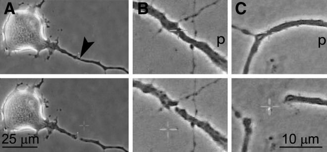
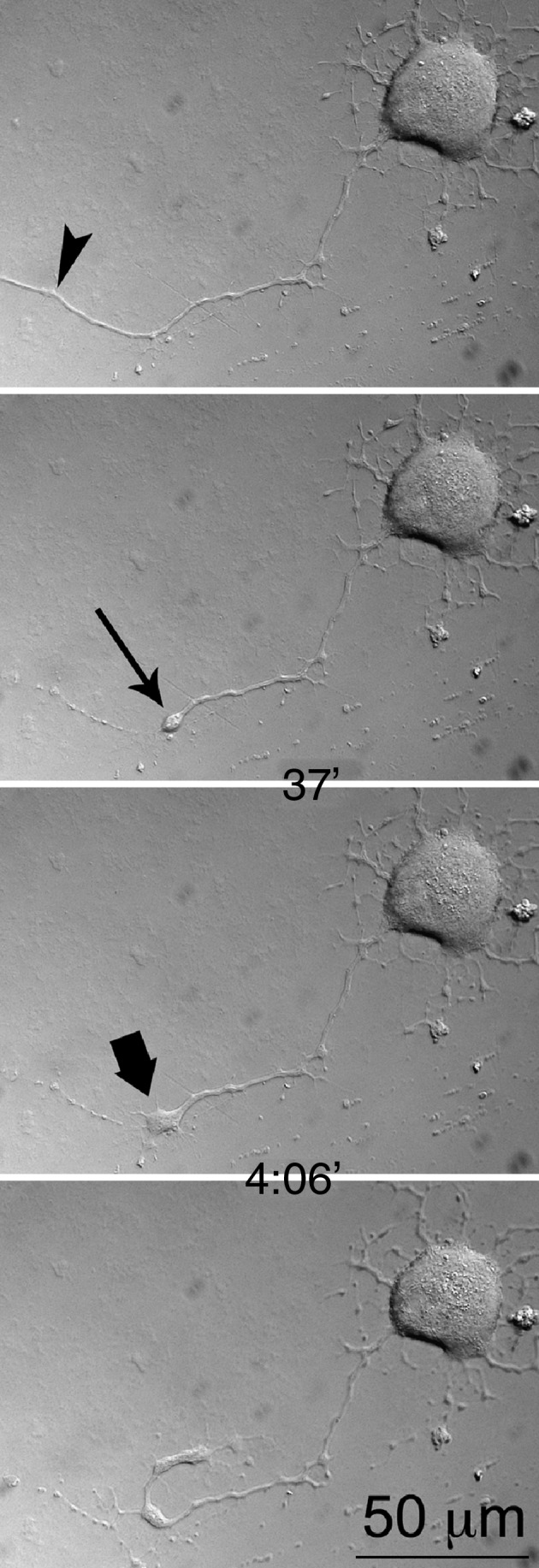
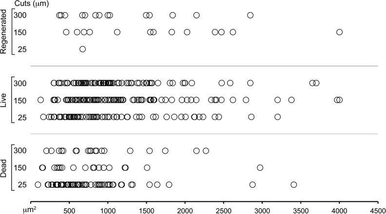
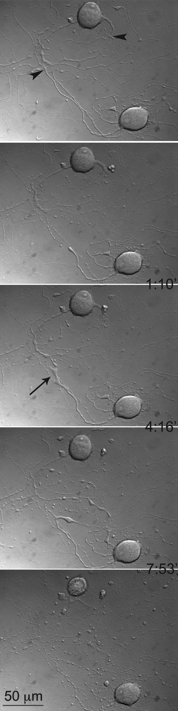
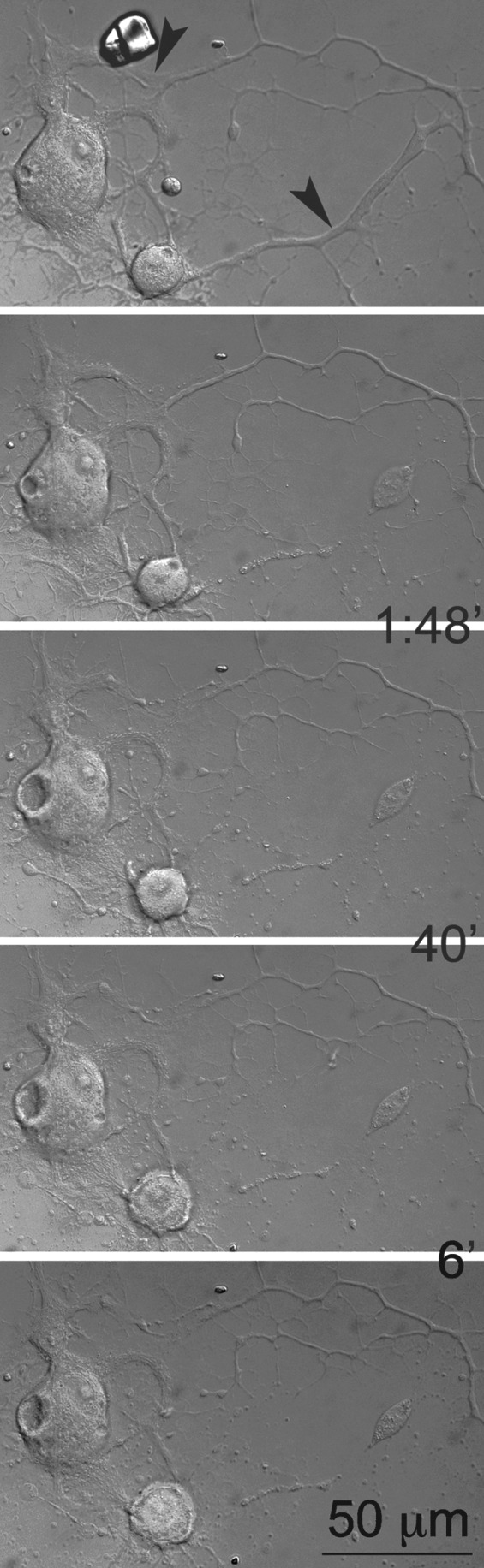
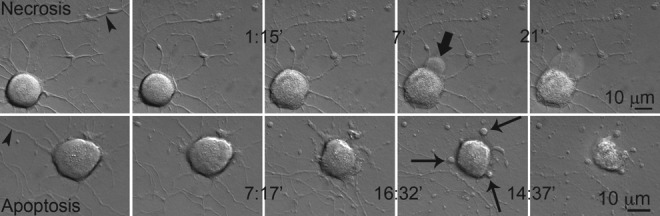
In order to quantify degenerative and regenerative changes and analyze the contribution of multiple factors to the outcome after neurite transection, we cultured adult mouse dorsal root ganglion neurons, and with a precise laser beam, we transected the nerve fibers they extended. Cell preparations were continuously visualized for 24 h with time-lapse microscopy. More distal cuts caused a more elongated field of degeneration, while thicker neurites degenerated faster than thinner ones. Transected neurites degenerated more if the uncut neurites of the same neuron simultaneously degenerated. If any of these uncut processes regenerated, the transected neurites underwent less degeneration. Regeneration of neurites was limited to distal cuts. Unipolar neurons had shorter regeneration than multipolar ones. Branching slowed the regenerative process, while simultaneous degeneration of uncut neurites increased it. Proximal lesions, small neuronal size, and extensive and rapid neurite degeneration were predictive of death of an injured neuron, which typically displayed necrotic rather than apoptotic form. In conclusion, this in vitro model proved useful in unmasking many new aspects and correlates of mechanically-induced neurite injury.
Figures
Similar articles
-
Calcium influx is necessary for optimal regrowth of transected neurites of rat sympathetic ganglion neurons in vitro.Neuroscience. 2001;102(4):945-57. doi: 10.1016/s0306-4522(00)00514-5. Neuroscience. 2001. PMID: 11182256
-
A critical role for macrophages near axotomized neuronal cell bodies in stimulating nerve regeneration.J Neurosci. 2013 Oct 9;33(41):16236-48. doi: 10.1523/JNEUROSCI.3319-12.2013. J Neurosci. 2013. PMID: 24107955 Free PMC article.
-
Modeling axonal injury in vitro: injury and regeneration following acute neuritic trauma.J Neurosci Methods. 2000 Oct 15;102(1):69-79. doi: 10.1016/s0165-0270(00)00282-x. J Neurosci Methods. 2000. PMID: 11000413
-
The Dorsal Column Lesion Model of Spinal Cord Injury and Its Use in Deciphering the Neuron-Intrinsic Injury Response.Dev Neurobiol. 2018 Oct;78(10):926-951. doi: 10.1002/dneu.22601. Epub 2018 May 11. Dev Neurobiol. 2018. PMID: 29717546 Free PMC article. Review.
-
Mechanisms of neurite repair.Curr Opin Neurobiol. 2020 Aug;63:53-58. doi: 10.1016/j.conb.2020.02.010. Epub 2020 Apr 8. Curr Opin Neurobiol. 2020. PMID: 32278210 Free PMC article. Review.
Cited by
-
Adult mouse dorsal root ganglia neurons form aberrant glutamatergic connections in dissociated cultures.PLoS One. 2021 Mar 3;16(3):e0246924. doi: 10.1371/journal.pone.0246924. eCollection 2021. PLoS One. 2021. PMID: 33657119 Free PMC article.
-
A Brief Review of In Vitro Models for Injury and Regeneration in the Peripheral Nervous System.Int J Mol Sci. 2022 Jan 13;23(2):816. doi: 10.3390/ijms23020816. Int J Mol Sci. 2022. PMID: 35055003 Free PMC article. Review.
-
Neuroprotective Effects of Curcumin-Loaded Emulsomes in a Laser Axotomy-Induced CNS Injury Model.Int J Nanomedicine. 2020 Nov 20;15:9211-9229. doi: 10.2147/IJN.S272931. eCollection 2020. Int J Nanomedicine. 2020. PMID: 33244233 Free PMC article.
-
An In Vitro Model for Conditioning Lesion Effect.Cell Mol Neurobiol. 2019 Jan;39(1):61-71. doi: 10.1007/s10571-018-0633-2. Epub 2018 Nov 10. Cell Mol Neurobiol. 2019. PMID: 30415355 Free PMC article.
-
Axon-specific microtubule regulation drives asymmetric regeneration of sensory neuron axons.Elife. 2025 Feb 24;13:RP104069. doi: 10.7554/eLife.104069. Elife. 2025. PMID: 39992313 Free PMC article.
References
-
- Waller A. Experiments on the section of the glossopharyngeal and hypoglossal nerves of the frog, and observations of the alterations produced thereby in the structure of their primitive fibres. Philos. Trans. R. Soc. Lond. B. Biol. Sci. 1850;140:423–429.
-
- Cavanagh J.B. The ‘dying back’ process. A common denominator in many naturally occurring and toxic neuropathies. Arch. Pathol. Lab. Med. 1979;103:659–664. - PubMed
-
- Fry E.J. Ho C. David S. A role for Nogo receptor in macrophage clearance from injured peripheral nerve. Neuron. 2007;53:649–662. - PubMed
Publication types
MeSH terms
LinkOut - more resources
Full Text Sources
