. 2010 Mar;201(5):783-92.
doi: 10.1086/650493.

Intracellular Mycobacterium avium intersect transferrin in the Rab11(+) recycling endocytic pathway and avoid lipocalin 2 trafficking to the lysosomal pathway

Affiliations

Intracellular Mycobacterium avium intersect transferrin in the Rab11(+) recycling endocytic pathway and avoid lipocalin 2 trafficking to the lysosomal pathway

Oyvind Halaas et al. J Infect Dis. 2010 Mar.

Abstract

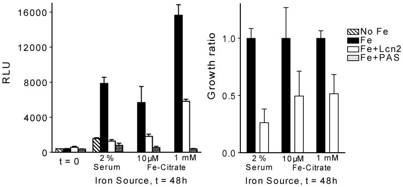
Iron is an essential nutrient for microbes, and many pathogenic bacteria depend on siderophores to obtain iron. The mammalian innate immunity protein lipocalin 2 (Lcn2; also known as neutrophil gelatinase-associated lipocalin, 24p3, or siderocalin) binds the siderophore carboxymycobactin, an essential component of the iron acquisition apparatus of mycobacteria. Here we show that Lcn2 suppressed growth of Mycobacterium avium in culture, and M. avium induced Lcn2 production from mouse macrophages. Lcn2 also had elevated levels and initially limited the growth of M. avium in the blood of infected mice but did not impede growth in tissues and during long-term infections. M. avium is an intracellular pathogen. Subcellular imaging of infected macrophages revealed that Lcn2 trafficked to lysosomes separate from M. avium, whereas transferrin was efficiently transported to the mycobacteria. Thus, mycobacteria seem to reside in the Rab11(+) endocytic recycling pathway, thereby retaining access to nutrition and avoiding endocytosed immunoproteins like Lcn2.


Conflict of interest statement

Potential conflicts of interest: none

Figures

Figure 1
Figure 1
Lcn2 suppresses growth of M. avium. Luciferase-expressing M. avium were grown in iron-free medium supplemented with serum (2 %) from Lcn2 KO mice or ferric-citrate (10 μM or 1 mM) as sources of iron, and Lcn2 (6 μg/ml) or p-aminosalicylic acid (PAS, 10 μg/ml). Growth was assessed by measuring luciferase activity in cell lysates (RLU). Data in the left panel are shown as mean RLU ± s.d. from one experiment. In the right panel the growth of M. avium in the presence of Lcn2 relative to growth in full-iron medium is shown as an average from repeated experiments.
Figure 2
Figure 2
Macrophages are induced to produce Lcn2. Day 7 mouse bone marrow derived macrophages (BMDM) were incubated with TLR-ligands (A) or infected with M. avium (B, C) and the supernatants assessed for Lcn2 by ELISA. (A) TLR-ligands (20 hours): 100 ng/ml Pam3CysSK4, 1 μg/ml LTA, 100 ng/ml FSL1, 20 μg/ml poly(I:C), 10 ng/ml LPS, 200 ng/ml flagellin, 5 μM R848, 5 μM CpG. (B) M. avium for indicated time and multiples of infection, MOI. (C) BMDM from WT, Lcn2 KO, MyD88 KO and TRIF KO infected with M. avium at MOIs as indicated for 4 days. All data are presented as means ± s.d. of triplicate measurements from one experiment repeated two (A) three (B) or one (C) times.
Figure 3
Figure 3
Macrophages endocytose Lcn2. (A) Accumulation of Lcn2. Day 7 BMDM were incubated with 5 μg/ml Lcn2-A488 and left for indicated times before analysis by flow cytometry. Data are represented as flow histogram (left) or mean fluorescence index of all time points (right). (B) Lcn2 is transported to lysosomes. Day 7 BMDM were incubated with 5 μg/ml Lcn2-A647 together with 5 μg/ml transferrin-A546 (Tf), 1 μg/ml Dextran-A488 (Dex), or 5 μg/ml RAP-A488 (RAP) for indicated times, or with 25 nM Lysotracker green (Lyso) for the last 30 minutes before analysis with confocal microscopy. Scale bars are 10 μm.
Figure 4
Figure 4
Effect of Lcn2 on M. avium growth in macrophages and in vivo in mice. BMDM from WT or Lcn2 KO mice were infected with (A) M. avium-luciferase for 4 days at MOI 3:1 and growth analyzed by luminescence (RLU), (B) M. avium-GFP for 4 days at MOI 3:1 and growth analyzed by FACS analysis (Mean Fluorescence Index), (C) M. avium for 4 days at MOI 3:1 and growth assayed by CFU. (D) Shows the effect of rLcn2 (8 μg/ml) on M. avium-luciferase growth in macrophages for 6 days. Data in (A-D) are shown as means ± s.d. from single experiments repeated two to ten times. Dose and kinetics from these experiments can be found in Supplementary figure S1. (E, F) WT and Lcn2-deficient mice were infected i.p. with 5 × 10⁷ CFU M. avium for indicated times before blood, spleen and liver were collected from 4-5 mice per group. Results from one experiment are shown. (E) Serum Lcn2 as measured by ELISA. Black squares represent uninfected mice. White and grey boxes show Lcn2 levels with s.d. from groups of infected mice (n = 4-5). (F) Bacterial load in blood, spleen and liver. Combined p-values obtained from a univariate analysis of variance of three separate experiments were p = 0.1 and p = 0.05 for blood counts day 1 and 2 post infection, respectively. Combined p-values > 0.1 for blood counts day 4 and 8, and for all tissue counts (spleen, liver) at all time points. (G) Bacterial load in blood (black bars), CD45+ blood leukocytes (hatched bars), and leukocyte-depleted plasma (dotted bars) from Lcn2 KO and WT mice infected with M. avium, 1 day post infection. Recovery (open bars) represents the sum of CFUs from leukocyte and plasma fractions. Data are showed as means ± s.d. from 5 mice.
Figure 5
Figure 5
M. avium co-localize with transferrin but not Lcn2 or lysosomal markers. (A) Day 7 BMDM were infected for 4 days with M. avium-GFP or CFP and incubated for 2 hours with 5 μg/ml Lcn2-A647 together with 5 μg/ml transferrin-A546, 1 μg/ml dextran-A488, 5 μg/ml RAP-A488 or 25 nM Lysotracker green (only included the last 30 minutes). Scale bars are 10 μm. (B) Surface rendering model created from the 3D (z-stack) images in the upper panel of (A) of the macrophage infected with M. avium (green) given Lcn2 (red) and transferrin (blue). Scale bar is 5 μm. (C) Transmission electron microscopy picture of day 7 BMDM infected with M. avium and pulsed with BSA gold particles for 1.5 hours. Gold particles were mainly found accumulated in presumably lysosomal compartments (Ly) that were in close proximity to the bacteria (asterisks). (D) Day 7 BMDM infected with M. avium-GFP were labeled with anti-Lcn2 Abs. Scale bar is 10 μm.
Figure 6
Figure 6
M. avium interact with the Rab11 recycling compartment. (A) BMDM infected for 4 days with M. avium-CFP were labeled with anti-Rab11a, anti-Rab11b, anti-Rab4a, anti-Rab5a or anti-transferrin receptor Abs. Upper panel shows an overview (scale bar 10 μm), lower panel the indicated close-up shots. (B) A 3D z-stack of an infected Rab11a-labeled macrophage is shown as a top-down slice, a 3D surface rendering model and the 3D co-localization-channel of M. avium and Rab11a with the view-point indicated in the left panel showing direct overlap between Rab11a-compartments and mycobacteria. Scale bar is 10 μm. (C) Our model suggesting that M. avium locates with the Rab11+ endocytic recycling compartment, thereby obtaining transferrin while avoiding Lcn2 and other endocytosed substances that traffic to lysosomes for degradation.

References

    1. Schaible UE, Kaufmann SH. Iron and microbial infection. Nat Rev Microbiol. 2004;2:946–953. - PubMed
    1. Ratledge C, Dover LG. Iron metabolism in pathogenic bacteria. Annu Rev Microbiol. 2000;54:881–941. - PubMed
    1. Goetz DH, Holmes MA, Borregaard N, Bluhm ME, Raymond KN, Strong RK. The neutrophil lipocalin NGAL is a bacteriostatic agent that interferes with siderophore-mediated iron acquisition. Mol Cell. 2002;10:1033–1043. - PubMed
    1. Holmes MA, Paulsene W, Jide X, Ratledge C, Strong RK. Siderocalin (Lcn 2) also binds carboxymycobactins, potentially defending against mycobacterial infections through iron sequestration. Structure. 2005;13:29–41. - PubMed
    1. Flo TH, Smith KD, Sato S, et al. Lipocalin 2 mediates an innate immune response to bacterial infection by sequestrating iron. Nature. 2004;432:917–921. - PubMed

Publication types

MeSH terms