. 2010 Feb 1:7:6.
doi: 10.1186/1742-4690-7-6.

The mutation T477A in HIV-1 reverse transcriptase (RT) restores normal proteolytic processing of RT in virus with Gag-Pol mutated in the p51-RNH cleavage site

Affiliations

The mutation T477A in HIV-1 reverse transcriptase (RT) restores normal proteolytic processing of RT in virus with Gag-Pol mutated in the p51-RNH cleavage site

Michael E Abram et al. Retrovirology. .

Abstract

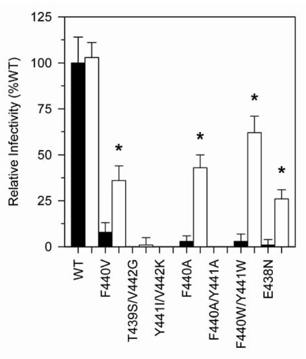
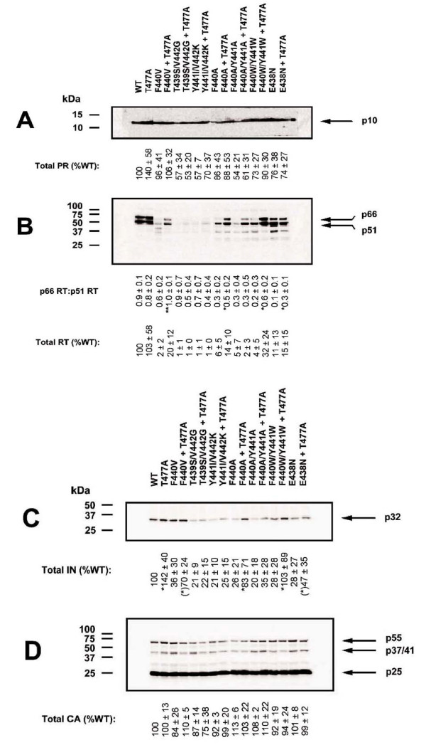
Background: The p51 subunit of the HIV-1 reverse transcriptase (RT) p66/p51 heterodimer arises from proteolytic cleavage of the RT p66 subunit C-terminal ribonuclease H (RNH) domain during virus maturation. Our previous work showed that mutations in the RT p51 downward arrowRNH cleavage site resulted in virus with defects in proteolytic processing of RT and significantly attenuated infectivity. In some cases, virus fitness was restored after repeated passage of mutant viruses, due to reversion of the mutated sequences to wild-type. However, in one case, the recovered virus retained the mutated p51 downward arrowRNH cleavage site but also developed an additional mutation, T477A, distal to the cleavage site. In this study we have characterized in detail the impact of the T477A mutation on intravirion processing of RT.

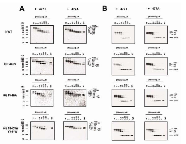
Results: While the T477A mutation arose during serial passage only with the F440V mutant background, introduction of this substitution into a variety of RT p51 downward arrowRNH cleavage site lethal mutant backgrounds was able to restore substantial infectivity and normal RT processing to these mutants. T477A had no phenotypic effect on wild-type HIV-1. We also evaluated the impact of T477A on the kinetics of intravirion Gag-Pol polyprotein processing of p51 downward arrowRNH cleavage site mutants using the protease inhibitor ritonavir. Early processing intermediates accumulated in p51 downward arrowRNH cleavage site mutant viruses, whereas introduction of T477A promoted the completion of processing and formation of the fully processed RT p66/p51 heterodimer.

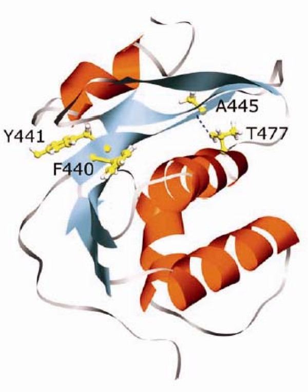
Conclusions: This work highlights the extraordinary plasticity of HIV-1 in adapting to seemingly lethal mutations that prevent RT heterodimer formation during virion polyprotein maturation. The ability of T477A to restore RT heterodimer formation and thus intravirion stability of the enzyme may arise from increased conformation flexibility in the RT p51 downward arrowRNH cleavage site region, due to loss of a hydrogen bond associated with the normal threonine residue, thereby enabling proteolytic cleavage near the normal RT p51 downward arrowRNH cleavage site.


Figures

Figure 1
Figure 1
Infectivity of WT and p51↓RNH ± T477A mutant virus in single cycle replication assays. HIV-1 derived from transfection of 293T cells was added to P4R5 indicator cells (5 × 10³), and infectivity was assessed 48 h post infection as indicated in Materials and Methods. Black and white bars indicate p51↓RNH - T477A or p51↓RNH + T477A mutant viruses respectively, derived from transfected 293T cells, normalized to 25 ng viral p24 at time of infection. Infectivity was determined after 48 h of culture by fluorescent measurement of β-galactosidase gene expression, as described in Materials and Methods. Data are means ± S.D. from 16 independent experiments. Asterisks (*) indicate statistical significance (p < 0.001) between -T477A and +T477A mutant viruses, calculated using a one-tailed Student's t-test assuming equal variance.
Figure 2
Figure 2
Effect of p51↓RNH ± T477A mutations on viral particle protein composition. Western blots of wild-type (WT) and p51↓RNH ± T477A mutant viruses (1 μg viral p24) generated by transfection of 293T cells and probed with (A) anti-PR, (B) anti-RT, (C) anti-IN, and (D) anti-p24 antibodies. The positions of molecular size markers are shown to the left of each panel. Arrows to the right of each panel indicate the positions and molecular masses of immunoreactive viral proteins. The relative mean proportion of p66 RT to p51 RT (p66:p51) and the total viral content of RT, IN and CA were determined from multiple experiments (n = 3) by densitometric scanning analysis of ECL-exposed blots under subsaturating conditions. Statistical significance of the T477A compensatory effect was determined for each individual mutant virus relative to its non-substituted counterpart using a one-tailed Student's t-test assuming equal variance. Asterisks indicate the degree of statistical significance in relation to the size of the type I error: (*)p < 0.10, *p < 0.05, **p < 0.01. In Figure 2A and 2B, WT and WT+T477A samples (two leftmost lanes) are from a different gel than the rest of the samples, as the number of wells in the electrophoresis apparatus was unable to accommodate all samples simultaneously. However, all electrophoresed samples had the same amount of p24 (see Methods) and were processed simultaneously (using two identical electrophoresis apparatus). Both resultant gels were imaged simultaneously by chemiluminescence as described in Materials and Methods.
Figure 3
Figure 3
Effect of p51↓RNH ± T477A mutations on ordered intravirion processing of Gag and Gag-Pol polyproteins. Virus-containing culture supernatants derived from the transfection of COS-7 cells in the presence of various concentrations of ritonavir were subjected to SDS-10% PAGE resolution and Western blotting analysis. (A) Pr160Gag-Pol and (B) Pr55gag polyprotein processing intermediates were visualized with anti-RT and anti-p24 monoclonal antibodies respectively, followed by ECL exposure. Analyses of p51↓RNH mutant viruses containing the wild-type 477T or the mutant 477A are in the left and right panels, respectively. The positions of molecular size markers are shown to the left of each panel. Lines to the right of each panel indicate the positions and estimated molecular masses of predicted polyprotein processing intermediates [24,27].
Figure 4
Figure 4
Positions of the p51↓RNH cleavage site and the residue T477 in HIV-1 RT. Ribbon diagram of amino acid residues 425-560 depicting the RNH domain of HIV-1 RT, adapted from PDB 1LDO[42]. The H-bond between the hydroxyl of T477 and the main chain of A445 is indicated by a dashed line. Details are provided in the text. The molecular graphics image was produced using the UCSF Chimera package from the resource for Biocomputing, Visualization, and Informatics at the University of California, San Francisco (supported by NIH P41 RR-01081) [43].

Similar articles

Cited by

References

    1. Tisdale M, Ertl P, Larder BA, Purifoy DJ, Darby G, Powell KL. Characterization of human immunodeficiency virus type 1 reverse transcriptase by using monoclonal antibodies: role of the C terminus in antibody reactivity and enzyme function. J Virol. 1988;62(10):3662–7. - PMC - PubMed
    1. Chattopadhyay D, Evans DB, Deibel MR Jr. Purification and characterization of heterodimeric human immunodeficiency virus type 1 (HIV-1) reverse transcriptase produced by in vitro processing of p66 with recombinant HIV-1 protease. J Biol Chem. 1992;267(20):14227–32. - PubMed
    1. Fan N, Rank KB, Leone JW. The differential processing of homodimers of reverse transcriptases from human immunodeficiency viruses type 1 and 2 is a consequence of the distinct specificities of the viral proteases. J Biol Chem. 1995;270(22):13573–9. - PubMed
    1. Hostomska Z, Matthews DA, Davies JF, Nodes BR, Hostomsky Z. Proteolytic release and crystallization of the RNase H domain of human immunodeficiency virus type 1 reverse transcriptase. J Biol Chem. 1991;266(22):14697–702. - PubMed
    1. Tomasselli AG, Sarcich JL, Barrett LJ, Reardon IM, Howe WJ, Evans DB, Sharma SK, Heinrikson RL. Human immunodeficiency virus type-1 reverse transcriptase and ribonuclease H as substrates of the viral protease. Protein Sci. 1993;2(12):2167–76. doi: 10.1002/pro.5560021216. - DOI - PMC - PubMed

Publication types

Substances

LinkOut - more resources