. 2010 Feb 1:9:12.
doi: 10.1186/1476-511X-9-12.

Docosahexaenoic acid prevents dendritic cell maturation and in vitro and in vivo expression of the IL-12 cytokine family

Affiliations

Docosahexaenoic acid prevents dendritic cell maturation and in vitro and in vivo expression of the IL-12 cytokine family

Weimin Kong et al. Lipids Health Dis. .

Abstract

Background: Acute and chronic inflammation play essential roles in inflammatory/autoimmune conditions. Protective anti-inflammatory effects of the n-3 fatty acids docosahexaenoic acid (DHA) and eicosapentaenoic acid (EPA) were reported in animal models of colitis, sepsis, and stroke. Since dendritic cells (DC) represent the essential cellular link between innate and adaptive immunity and have a prominent role in tolerance for self-antigens, we sought to investigate the impact of DHA on DC maturation and proinflammatory cytokine production.

Methods: Murine bone marrow-derived DC were treated with DHA and stimulated with various toll-like receptor (TLR) ligands. Flow cytometry was used to determine the levels of surface maturation markers and endocytic activity. Cytokine expression and secretion were measured by real-time RT-PCR and ELISA assays. PPARgamma and NFkappaB activity in nuclear extracts were determined by binding to specific oligonucleotide sequences using ELISA-based assays. In vivo effects of DHA were assessed in splenic DC from LPS-inoculated mice maintained on a DHA-enriched diet.

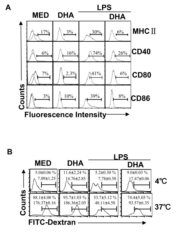
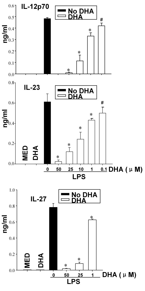
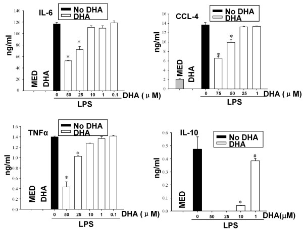
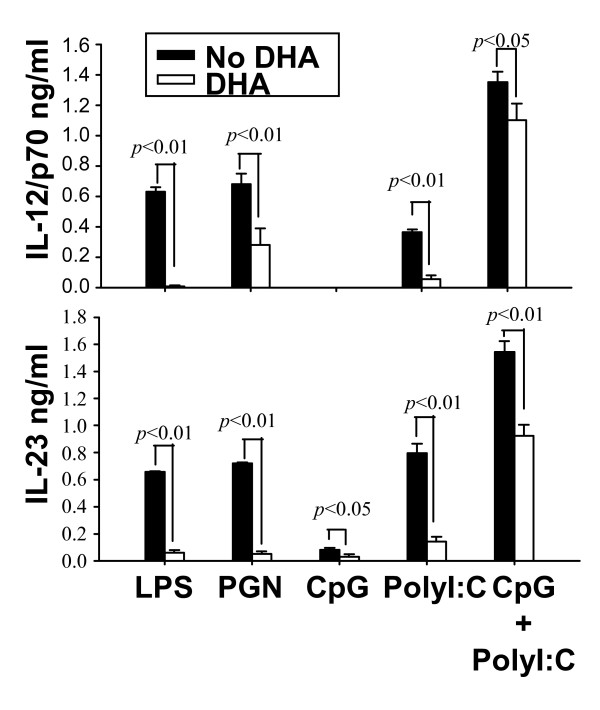
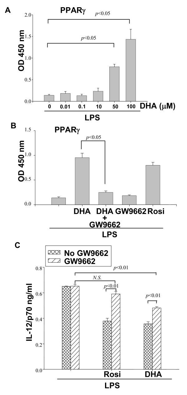
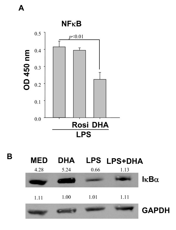
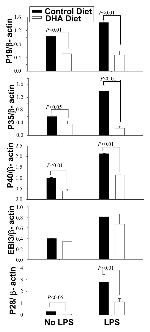
Results: DHA maintained the immature phenotype in bone marrow-derived DC by preventing the upregulation of MHCII and costimulatory molecules (CD40, CD80 and CD86) and maintaining high levels of endocytic activity. DHA inhibited the production of pro-inflammatory cytokines, including the IL-12 cytokine family (IL-12p70, IL-23, and IL-27), from DC stimulated with TLR2, 3, 4, and 9 ligands. DHA inhibition of IL-12 expression was mediated through activation of PPARgamma and inhibition of NFkappaBp65 nuclear translocation. DHA exerted a similar inhibitory effect on IL-12 and IL-23 expression in vivo in LPS-inoculated mice maintained on a DHA-enriched diet.

Conclusions: Exposure of bone marrow-derived DC to DHA resulted in the maintenance of an immature phenotype and drastic reduction in proinflammatory cytokine release. DHA inhibited the expression and secretion of the IL-12 cytokine family members (IL-12p70, IL-23 and IL-27), which play essential roles in the differentiation of the proinflammatory Th1/Th17 effector cells. The effect of DHA on IL-12 expression was mediated through activation of PPARgamma and inhibition of NFkappaB. Inhibition of IL-12 and IL-23 expression was also evident in splenic DC from mice fed a DHA-enriched diet, suggesting that dietary DHA acts as an anti-inflammatory agent in vivo.


Figures

Figure 1
Figure 1
DHA maintains DC immature phenotype and high endocytic capacity. CD11c+ DC (1 × 10⁶/ml) were treated with 50 μM DHA for 24 h, followed by LPS (0.1 μg/ml) for another 24 h. (A) Cells were analyzed for MHCII, CD40, CD80 and CD86 expression by flow cytometry. One representative experiment of three is shown. (B) DC were treated as in (A) and 5 × 10⁵ cells were incubated with FITC-dextran (0.5 mg/ml) at 4°C (negative control) or 37°C and assessed for endocytosis by flow cytometry. Results are expressed as percentage positive cells and geometric mean fluorescence (GMF) +/- SD.
Figure 2
Figure 2
DHA prevents expression of IL-12 family cytokines in LPS-treated DC. DC (1 × 10⁶/ml for IL-12p70 and IL-27 and 2 × 10⁶/ml for IL-23) were cultured in the presence of different concentration of DHA for 24 h, followed by LPS for 12 h (for IL-23) or 24 h (for IL-12p70 and IL-27). Supernatants were subjected to ELISA. Data represent the mean +/- SD. * p < 0.01, #p < 0.05, compared with LPS-treated samples (no DHA).
Figure 3
Figure 3
DHA prevents cytokine production in LPS-treated DC. CD11c+ DC (2 × 10⁶/ml for IL-10 or 1 × 10⁶/ml for TNFα, IL-6 and CCL-4) were treated with various concentrations of DHA for 24 h followed by LPS (0.1 μg/ml) for an additional 24 h. Supernatants were harvested and assayed for IL-6, CCL-4, IL-10 and TNFα by ELISA. Two independent cultures were tested in triplicate. Results are expressed as mean +/- SD. * p < 0.01, #p < 0.05, compared to LPS-treated samples (no DHA).
Figure 4
Figure 4
Effects of DHA on IL-12p70 production initiated by signaling through various TLRs. CD11c+ DC (1 × 10⁶ cells/ml for IL-12p70 and 2 × 10⁶ cells/ml for IL-23) were pretreated with 25 μM DHA for 24 h followed by different TLR ligands (LPS 0.1 μg/ml, PGN 10 μg/ml, CpG 1 μM, poly I:C 50 μg/ml, CpG 1 μM + poly I:C 50 μg/ml) for an additional 12 h (for IL-23) or 24 h (for IL-12p70). Supernatants were harvested and assayed for IL-12p70 or IL-23 by ELISA.
Figure 5
Figure 5
DHA activates PPARγ. (A) CD11c+ DC (5 × 10⁶) were pretreated with increasing concentrations of DHA for 24 h, followed by 0.1 μg/ml LPS stimulation for 16 h. Activation of nuclear PPARγ was evaluated through binding to oligonucleotides containing PPRE as described in Methods. (B) DC were pretreated with the PPARγ inhibitor GW9662 (10 μM) for 2 h, followed by 50 μM DHA or 1 μM of the specific PPARγ agonist Rosiglitazone (Rosi) for 24 h, and LPS (0.1 μg/ml) for an additional 16 h. Activation of PPARγ was determined as described in (A). (C) CD11c+ DC (1 × 10⁶/ml) were pretreated with 4 μM GW9662 for 2 h before the addition of 5 μM DHA or 0.4 μM Rosi (for 24 h) and LPS for an additional 24 h. Supernatants were collected and IL-12p70 production was determined by ELISA.
Figure 6
Figure 6
DHA blocks NFκB p65 nuclear translocation. (A) 1 × 10⁷DC were preincubated with (50 μM) DHA or (1 μM) Rosi for 24 h, followed by LPS treatment for 1 h. Presence of NFκB p65 in DC nuclear extracts was determined by binding to κB containing oligonucleotides. Results are expressed as relative activity, i.e. absorbance values above those observed in the absence of LPS stimulation. (B) DC were pretreated with DHA for 24 h, followed by LPS stimulation for 40 min, and cell lysates were subjected to Western blot analysis for IκBα. Numbers represent relative densitometric levels. One experiment out of three is shown.
Figure 7
Figure 7
DHA prevents in vivo expression of IL-12 family cytokines. C57BL/6 mice were maintained for 5 wks on regular or DHA-enriched diet. Mice were injected i.p. with LPS (50 μg per mouse) and 12 h later spleen CD11c+ DC were purified. Expression of p40, p35, p19, p28 and EBI3 was determined by real time RT-PCR.

Similar articles

Cited by

References

    1. Calder PC. The relationship between the fatty acid composition of immune cells and their function. Prostaglandins Leukot Essent Fatty Acids. 2008;79(3-5):101–108. doi: 10.1016/j.plefa.2008.09.016. - DOI - PubMed
    1. Chapkin RS, Kim W, Lupton JR, McMurray DN. Dietary docosahexaenoic and eicosapentaenoic acid: emerging mediators of inflammation. Prostaglandins Leukot Essent Fatty Acids. 2009;81(2-3):187–191. doi: 10.1016/j.plefa.2009.05.010. - DOI - PMC - PubMed
    1. Das UN. Essential fatty acids: biochemistry, physiology and pathology. Biotechnol J. 2006;1(4):420–439. doi: 10.1002/biot.200600012. - DOI - PubMed
    1. Orr SK, Bazinet RP. The emerging role of docosahexaenoic acid in neuroinflammation. Curr Opin Investig Drugs. 2008;9(7):735–743. - PubMed
    1. Serhan CN. Resolution phase of inflammation: novel endogenous anti-inflammatory and proresolving lipid mediators and pathways. Annu Rev Immunol. 2007;25:101–137. doi: 10.1146/annurev.immunol.25.022106.141647. - DOI - PubMed

Publication types