. 2010 Feb 1:10:29.
doi: 10.1186/1471-2407-10-29.

Pentastatin-1, a collagen IV derived 20-mer peptide, suppresses tumor growth in a small cell lung cancer xenograft model

Affiliations

Pentastatin-1, a collagen IV derived 20-mer peptide, suppresses tumor growth in a small cell lung cancer xenograft model

Jacob E Koskimaki et al. BMC Cancer. .

Abstract

Background: Angiogenesis is the formation of neovasculature from a pre-existing vascular network. Progression of solid tumors including lung cancer is angiogenesis-dependent. We previously introduced a bioinformatics-based methodology to identify endogenous anti-angiogenic peptide sequences, and validated these predictions in vitro in human umbilical vein endothelial cell (HUVEC) proliferation and migration assays.

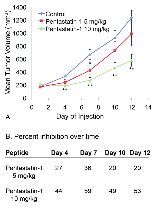
Methods: One family of peptides with high activity is derived from the alpha-fibrils of type IV collagen. Based on the results from the in vitro screening, we have evaluated the ability of a 20 amino acid peptide derived from the alpha5 fibril of type IV collagen, pentastatin-1, to suppress vessel growth in an angioreactor-based directed in vivo angiogenesis assay (DIVAA). In addition, pentastatin-1 suppressed tumor growth with intraperitoneal peptide administration in a small cell lung cancer (SCLC) xenograft model in nude mice using the NCI-H82 human cancer cell line.

Results: Pentastatin-1 decreased the invasion of vessels into angioreactors in vivo in a dose dependent manner. The peptide also decreased the rate of tumor growth and microvascular density in vivo in a small cell lung cancer xenograft model.

Conclusions: The peptide treatment significantly decreased the invasion of microvessels in angioreactors and the rate of tumor growth in the xenograft model, indicating potential treatment for angiogenesis-dependent disease, and for translational development as a therapeutic agent for lung cancer.


Figures

Figure 1
Figure 1
In vitro cell viability assay. A. NCI-H82 small cell lung cancer cells (H82) and 3T3 fibroblasts (3T3) were plated at 2,000 cells/well. Pentastatin-1 peptide was applied at increasing concentrations up to 100 μg/mL, and incubated for three days. Cell viability was determined using the WST-1 colorimetric agent, and scaled to the untreated experimental control. Differences between H82 and 3T3 treatments are statistically significant as determined by the Student's t-test at p < 0.05 * and p < 0.01 **. B. BrdU incorporation for NCI-H82 small cell lung cancer cells and 3T3 fibroblasts. Cells were plated at 10,000 cells/well with pentastatin-1 at concentrations of 3.8, 15 and 60 μg/mL in the presence of a BrdU label for 24 hours. BrdU incorporation was detected by the BrdU-antibody peroxidase conjugate as a measurement for cell proliferation by DNA synthesis. Pentastatin-1 decreases cell proliferation by 50% for H82 and 33% for 3T3 at 60 μg/mL. Statistical significance is determined at p < 0.05 * and p < 0.01 ** by Student's t-test.
Figure 2
Figure 2
angioreactor-based in vivo assay. A. Directed in vivo angiogenesis assay (DIVAA) showing silicone tubes with and without application of pentastatin-1 at 200 μg/mL. Angioreactors were implanted subcutaneously into the abdominal region of C57BL/6 mice for 12 days, and vessels allowed to infiltrate. B. Peptides were mixed at concentrations of 30, 50, 100, and 200 μg/mL with Matrigel (BD Biosciences, San Jose, CA), and vascularization assayed and scaled where 100% represents the mean of the untreated controls (Ctrl). Results are significantly less than controls at p < 0.05 *.
Figure 3
Figure 3
NCI-H82 small cell lung cancer xenograft in nude mice. A. Mean tumor volume reported over time for the NCI-H82 SCLC xenograft model. Peptides were administered intraperitoneally, once per day, at two separate concentrations of 5 mg/kg or 10 mg/kg in comparison to a PBS-treated control and a scrambled peptide equivalent (not shown). Pentastatin-1 significantly suppressed growth for all days at 10 mg/kg by Student's t-test at p < 0.01 ** and demonstrated a dose response in vivo. B. Percent inhibition from day 1 reported over time for pentastatin-1 at 5 mg/kg and 10 mg/kg.
Figure 4
Figure 4
CD31 antibody staining for microvascular density. A. Immunohistochemistry showing CD31 as a marker for endothelial cells, and caspase-3 antibody staining for apoptotic cells in the control, and pentastatin-1 at 5 mg/kg and 10 mg/kg as a marker for endothelial cells and blood vessel density. B. Quantification of microvascular density using CD31 antibody. Blood vessels are stained in tumor sections using the CD31 antibody, and endothelial cells quantified by pixel intensity representing the quantity of endothelial cells as a percentage of the control. Three sample tumors for each condition with eight cross sections were quantified. Pentastatin-1 at 5 mg/kg and 10 mg/kg showed a statistically significant 23% and 24% decrease in microvascular density, respectively, from the control at p < 0.05 *. C. Quantification of the mean number of apoptotic cells per frame at 20× magnification for the PBS-treated control, and pentastatin-1 at 5 mg/kg 10 mg/kg. Controls show an average 7 cells/frame, while pentastatin-1 at 5 mg/kg shows 19 cells/frame and pentastatin-1 at 10 mg/kg show an average of 22 cells/frame. Results are statistically significant at p < 0.01**.

References

    1. Folkman J. Tumor angiogenesis: therapeutic implications. N Engl J Med. 1971;285(21):1182–1186. - PubMed
    1. Savai R, Langheinrich AC, Schermuly RT, Pullamsetti SS, Dumitrascu R, Traupe H, Rau WS, Seeger W, Grimminger F, Banat GA. Evaluation of angiogenesis using micro-computed tomography in a xenograft mouse model of lung cancer. Neoplasia. 2009;11(1):48–56. - PMC - PubMed
    1. Jemal A, Siegel R, Ward E, Hao Y, Xu J, Murray T, Thun MJ. Cancer statistics, 2008. CA Cancer J Clin. 2008;58(2):71–96. doi: 10.3322/CA.2007.0010. - DOI - PubMed
    1. Goudar RK, Vlahovic G. Hypoxia, angiogenesis, and lung cancer. Curr Oncol Rep. 2008;10(4):277–282. doi: 10.1007/s11912-008-0043-6. - DOI - PubMed
    1. Vokes E, Herbst R, Sandler A. Angiogenesis inhibition in the treatment of lung cancer. Clin Adv Hematol Oncol. 2006;4(11 Suppl 23):1–10. quiz 11-12. - PubMed

Publication types

MeSH terms