. 2010 Feb 2:9:25.
doi: 10.1186/1476-4598-9-25.

Axin downregulates TCF-4 transcription via beta-catenin, but not p53, and inhibits the proliferation and invasion of lung cancer cells

Affiliations

Axin downregulates TCF-4 transcription via beta-catenin, but not p53, and inhibits the proliferation and invasion of lung cancer cells

Lian-He Yang et al. Mol Cancer. .

Abstract

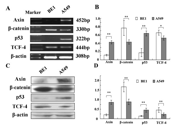
Background: We previously reported that overexpression of Axin downregulates T cell factor-4 (TCF-4) transcription. However, the mechanism(s) by which Axin downregulates the transcription and expression of TCF-4 is not clear. It has been reported that beta-catenin promotes and p53 inhibits TCF-4 transcription, respectively. The aim of this study was to investigate whether beta-catenin and/or p53 is required for Axin-mediated downregulation of TCF-4.

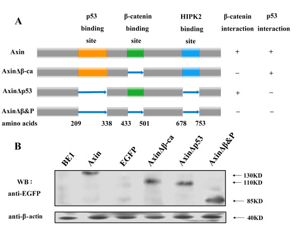
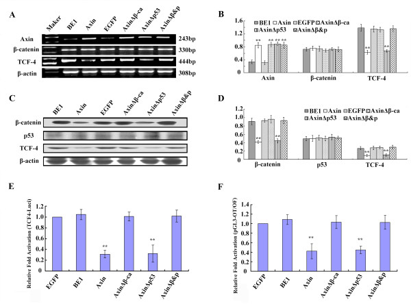
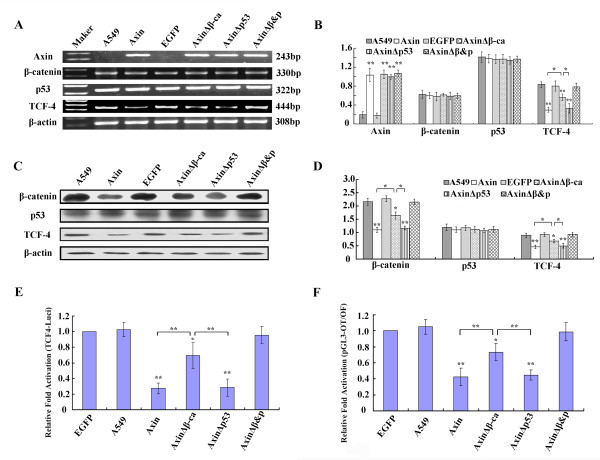
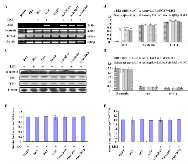
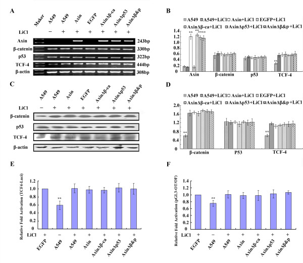
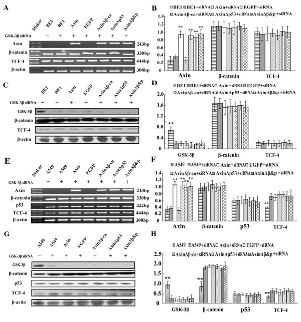
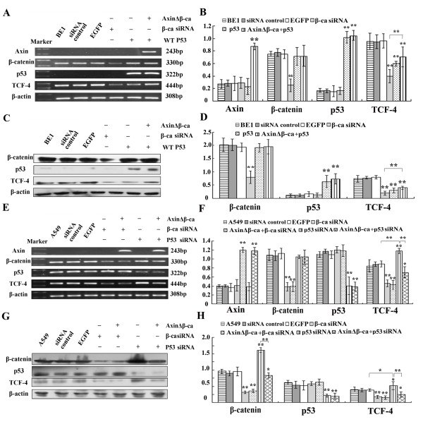
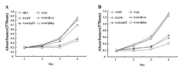
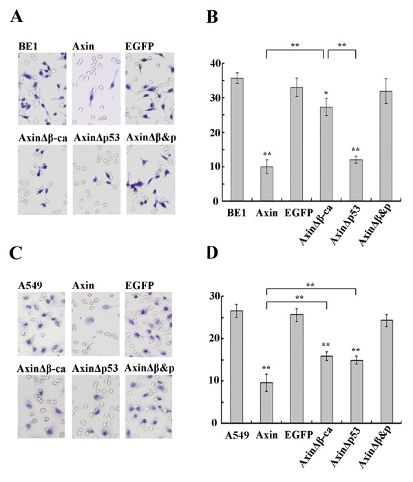
Results: Axin mutants that lack p53/HIPK2 and/or beta-catenin binding domains were expressed in lung cancer cells, BE1 (mutant p53) and A549 (wild type p53). Expression of Axin or AxinDeltap53 downregulates beta-catenin and TCF-4, and knock-down of beta-catenin upregulates TCF-4 in BE1 cells. However, expression of AxinDeltabeta-ca into BE1 cells did not downregulate TCF-4 expression. These results indicate that Axin downregulates TCF-4 transcription via beta-catenin. Although overexpression of wild-type p53 also downregulates TCF-4 in BE1 cells, cotransfection of p53 and AxinDeltabeta-ca did not downregulate TCF-4 further. These results suggest that Axin does not promote p53-mediated downregulation of TCF-4. Axin, AxinDeltap53, and AxinDeltabeta-ca all downregulated beta-catenin and TCF-4 in A549 cells. Knock-down of p53 upregulated beta-catenin and TCF-4, but cotransfection of AxinDeltabeta-ca and p53 siRNA resulted in downregulation of beta-catenin and TCF-4. These results indicate that p53 is not required for Axin-mediated downregulation of TCF-4. Knock-down or inhibition of GSK-3beta prevented Axin-mediated downregulation of TCF-4. Furthermore, expression of Axin and AxinDeltap53, prevented the proliferative and invasive ability of BE1 and A549, expression of AxinDeltabeta-ca could only prevented the proliferative and invasive ability effectively.

Conclusions: Axin downregulates TCF-4 transcription via beta-catenin and independently of p53. Axin may also inhibits the proliferation and invasion of lung cancer cells via beta-catenin and p53.


Figures

Figure 1
Figure 1
The mRNA and protein expression levels of Axin and other factors in BE1 and A549 cell line. (A) RT-PCR analysis of BE1 and A549 cells using the indicated specific primers. (B) Quantitative representation of the RT-PCR analysis. (C) Western blot analyses of BE1 and A549 cells using specific antibodies to the indicated proteins. (D) Quantitative representation of the Western blot anlaysis. * P < 0.05, ** P < 0.01.
Figure 2
Figure 2
The composition and expression of Axin mutants. (A) Schematic representation of the mutants of the Axin mutants used in this study. The binding sites for HIPK2, β-catenin and p53 are indicated. (B) BE1 cells were transfected with the indicated EGFP-tagged Axin construct. Total cell lysates were resolved by SDS-PAGE and Western blot analysis was performed using anti-EGFP antibody to detect the expression of the different Axin mutants.
Figure 3
Figure 3
The effect of transfecting various Axin plasmids into BE1 cells. Axin, AxinΔβ-ca, AxinΔp53 and AxinΔβ&P were transfected into BE1 cells. (A) Forty-eight hours after transfection total RNA was isolated and RT-PCR analysis was performed using the indicated primers. (B) Quantification of mRNA expression after transfection. (C) Proteins expression of BE1 cells 48 h after transfection using the indicated antibodies. (D) Quantification of protein expression 48 h after transfection of BE1 cells with (E) the TCF-4 promoter construct and (F) TCF reporter construct. * P < 0.05, ** P < 0.01.
Figure 4
Figure 4
The effect of transfecting various Axin plasmids into A549 cells. Axin, AxinΔβ-ca, AxinΔp53 and AxinΔβ&P were transfected into A549 cells. (A) Forty-eight hours after transfection total RNA was isolated and RT-PCR analysis was performed using the indicated primers. (B) Quantification of mRNA expression after transfection. (C) Protein expression of A549 cells 48 h after transfection using the indicated antibodies. (D) Quantification of protein expression 48 h after transfection of A549 cells with (E) the TCF-4 promoter construct and (F) TCF reporter construct. * P < 0.05, ** P < 0.01.
Figure 5
Figure 5
The effect of LiCl treatment on BE1 cells transfected with different Axin plasmids. Axin, AxinΔβ-ca, AxinΔp53 and AxinΔβ&P were transfected into BE1 cells. Twenty-four hours post-transfection, cells were treated with LiCl (20 mM). (A) Forty-eight hours after transfection total RNA and RT-PCR analysis was performed using the indicated primers. (B) Quantification of mRNA expression after transfection. (C) Protein expression in BE1 cells 48 h after transfection using the indicated antibodies. (D) Quantification of protein expression 48 h after transfection with (E) the TCF-4 promoter construct and (F) TCF reporter construct. * P < 0.05, ** P < 0.01.
Figure 6
Figure 6
The effect of LiCl treatment on A549 cells transfected with different Axin plasmids. Axin, AxinΔβ-ca, AxinΔp53 and AxinΔβ&P were transfected into A549 cells. Twenty-four hours post-transfection, cells were treated with LiCl (20 mM). (A) Forty-eight hours after transfection total RNA and RT-PCR analysis was performed using the indicated primers. (B) Quantification of mRNA expression after transfection. (C) Protein expression in A549 cells 48 h after transfection using the indicated antibodies. (D) Quantification of protein expression 48 h after transfection with (E) the TCF-4 promoter construct and (F) TCF reporter construct. * P < 0.05, ** P < 0.01.
Figure 7
Figure 7
The effect of GSK-3β siRNA treatment on BE1 cells and A549 cells transfected with different Axin plasmids. Axin, AxinΔβ-ca, AxinΔP53 and AxinΔβ&P were cotransfected with GSK-3? siRNA into BE1 (A-D) and A549 (E-H) cells. (A&E) Forty-eight hours after transfection total RNA and RT-PCR analysis was performed using the indicated primers. (B&F) Quantification of mRNA expression after cotransfection. (C&G) Western blot analysis of BE1 and A549 cells 48 h after transfection using the indicated antibodies. (D&H) Quantification of protein expression after transfection. * P < 0.05, ** P < 0.01.
Figure 8
Figure 8
Axin-mediated downregulation of TCF-4 is p53-independent. BE1 cells were transfected with wild-type p53 or β-catenin siRNA, respectively, cotransfection of wild-type p53 and AxinΔβ-ca was also performed in BE1 cells. A549 cells were transfected with p53 siRNA or β-catenin siRNA, respectively, and cotransfection of β-catenin siRNA and AxinΔβ-ca, p53 siRNA and AxinΔβ-ca were also performed in A549 cells. (A&E) Forty-eight hours after transfection total RNA and RT-PCR analysis was performed using the indicated primers. EGFP and control siRNA served as control groups. (B&F) Quantification of mRNA expression after transfection. (C&G) Western blot analysis was performed 48 h after transfection using the indicated antibodies. (D&H) Quantification of protein expression after transfection * P < 0.05, ** P < 0.01.
Figure 9
Figure 9
Transfecting various Axin plasmids influences the proliferative ability of BE1 and A549 cells. Cells were transfected with Axin, AxinΔβ-ca, AxinΔP53 and AxinΔβ&P and plated into wells 12 h post-transfection. MTT assay was performed every day for four days. Growth curves were generated for BE1 cells (A) and A549 cells (B).
Figure 10
Figure 10
Expression of various Axin constructs decreases the invasive ability of BE1 and A549 cells. Cells were seeded on the upper chamber (5 × 10⁴ cells/well) 24 h after transfection and incubated for 48 h. (A&C) A representative microscope field of filters under the Matrigel from BE1 and A549, respectively. (B&D) Quantification of the number of migrated BE1 and A549 cells, respectively. * P < 0.05, ** P < 0.01.

Similar articles

Cited by

References

    1. Lustig B, Behrens J. The Wnt signaling pathway and its role in tumor development. J Cancer Res Clin Oncol. 2003;129:199–221. - PubMed
    1. Ueta T, Ikeguchi M, Hirooka Y, Kaibara N, Terada T. Beta-catenin and cyclin D1 expression in human hepatocellular carcinoma. Oncol Rep. 2002;9:1197–1203. - PubMed
    1. Gerstein AV, Almeida TA, Zhao G, Chess E, Shih IeM, Buhler K, Pienta K, Rubin MA, Vessella R, Papadopoulos N. APC/CTNNB1 (beta-catenin) pathway alterations in human prostate cancers. Genes Chromosomes Cancer. 2002;34:9–16. doi: 10.1002/gcc.10037. - DOI - PubMed
    1. Cheng XX, Sun Y, Chen XY, Zhang KL, Kong QY, Liu J, Li H. Frequent translocalization of beta-catenin in gastric cancers and its relevance to tumor progression. Oncol Rep. 2004;11:1201–1207. - PubMed
    1. Xu HT, Wang L, Lin D, Liu Y, Liu N, Yuan XM, Wang EH. Abnormal beta-catenin and reduced axin expression are associated with poor differentiation and progression in non-small cell lung cancer. Am J Clin Pathol. 2006;125:534–541. - PubMed

Publication types

MeSH terms