. 2010 Feb 2:8:11.
doi: 10.1186/1741-7007-8-11.

Adherens junctions connect stress fibres between adjacent endothelial cells

Affiliations

Adherens junctions connect stress fibres between adjacent endothelial cells

Jaime Millán et al. BMC Biol. .

Abstract

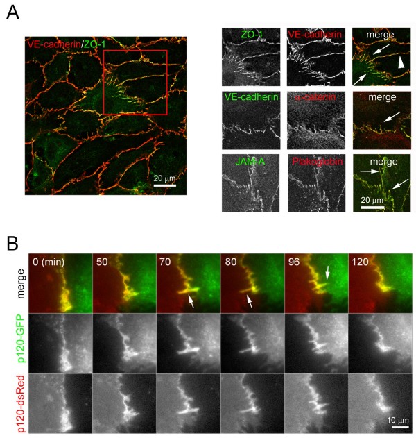
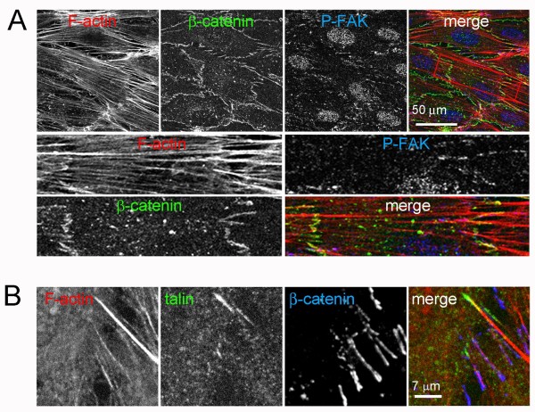
Background: Endothelial cell-cell junctions maintain endothelial integrity and regulate vascular morphogenesis and homeostasis. Cell-cell junctions are usually depicted with a linear morphology along the boundaries between adjacent cells and in contact with cortical F-actin. However, in the endothelium, cell-cell junctions are highly dynamic and morphologically heterogeneous.

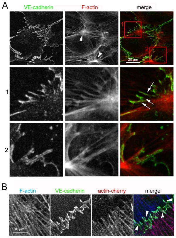
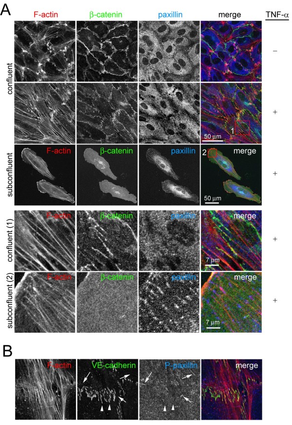
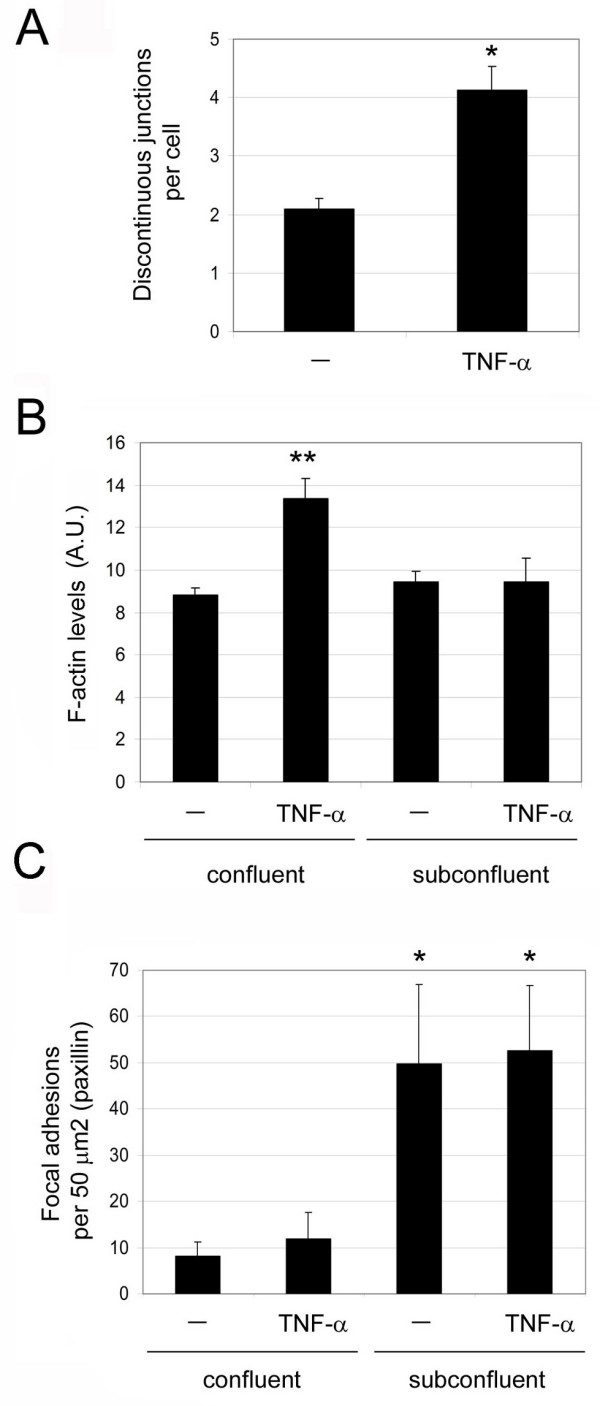
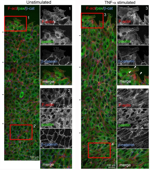
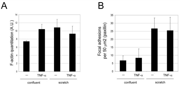
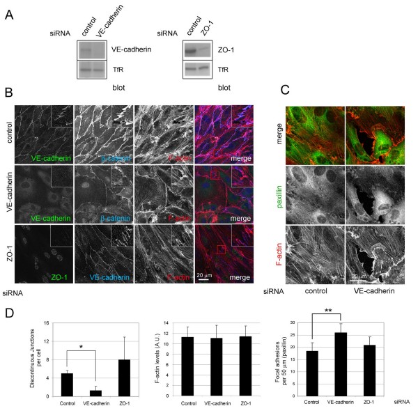
Results: We report that endothelial cell-cell junctions can attach to the ends of stress fibres instead of to cortical F-actin, forming structures that we name discontinuous adherens junctions (AJ). Discontinuous AJ are highly dynamic and are increased in response to tumour necrosis factor (TNF)-alpha, correlating with the appearance of stress fibres. We show that vascular endothelial (VE)-cadherin/beta-catenin/alpha-catenin complexes in discontinuous AJ are linked to stress fibres. Moreover, discontinuous AJ connect stress fibres from adjacent cells independently of focal adhesions, of which there are very few in confluent endothelial cells, even in TNF-alpha-stimulated cells. RNAi-mediated knockdown of VE-cadherin, but not zonula occludens-1, reduces the linkage of stress fibres to cell-cell junctions, increases focal adhesions, and dramatically alters the distribution of these actin cables in confluent endothelial cells.

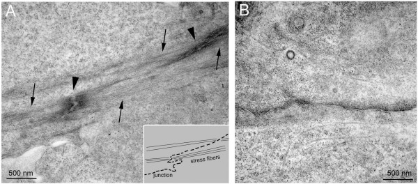
Conclusions: Our results indicate that stress fibres from neighbouring cells are physically connected through discontinuous AJ, and that stress fibres can be stabilized by AJ-associated multi-protein complexes distinct from focal adhesions.


Figures

Figure 1
Figure 1
Composition of discontinuous adherens junction (AJ). (A) Human umbilical vein endothelial cells (HUVECs) were grown at confluency for 72 h in EGM-2 growth medium, fixed, permeabilized and stained with antibodies to the indicated junctional proteins. Top right panels show single staining of the merged image on the left. Arrows indicate discontinuous cell-cell junctions; arrowhead, linear cell-cell junctions. (B) HUVECs were nucleofected with plasmids coding for p120-catenin-green fluorescent protein (p120-GFP) or p120-catenin-red fluorescent protein (p120-dsRed), cells from the two transfections were mixed and plated at confluence for 24 h in growth medium. Cell images were acquired by fluorescence time-lapse microscopy, 1 frame/min. Representative images are shown. Arrows show discontinuous AJ.
Figure 2
Figure 2
Discontinuous adherens junction (AJ) anchor stress fibres at cell borders. (A) Confluent human umbilical vein endothelial cells (HUVECs) in EGM-2 growth medium were stained for actin filaments (F-actin; TRITC-phalloidin) and vascular endothelial (VE)-cadherin. Bottom panels are enlargements of red boxed areas from top panel that are enriched in discontinuous AJ. (B) HUVECs were nucleofected with a plasmid encoding actin-cherry and plated at confluence for 24 h to 48 h in growth medium. Cells were fixed and stained for VE-cadherin or F-actin. Arrowheads indicate discontinuous AJ.
Figure 3
Figure 3
Electron microscopy analysis of endothelial cell-cell borders. Confluent human umbilical vein endothelial cells (HUVECs) either (B) unstimulated or (A) tumour necrosis factor-α-stimulated for 18 h were fixed and processed for electron microscopy. Arrows denote actin filament bundles, whilst arrowheads indicate position of electron-dense junctional structures. Inset: cartoon of panel A illustrating placement of stress fibres with respect to junctional contacts, confirming a connection between junctions and actin filaments, which is not observed at linear junctions (B).
Figure 4
Figure 4
Effect of tumour necrosis factor (TNF)-α and confluence on discontinuous adherens junction (AJ), stress fibres and focal adhesions. (A) Effect of TNF-α on actin filament, β-catenin and paxillin distribution. Confluent human umbilical vein endothelial cells (HUVECs) were starved for 4 h and then, as indicated, stimulated with 10 ng/ml TNF-α for 20 h. In parallel, subconfluent HUVECs were also starved and stimulated with TNF-α. Cells were fixed and distribution of F-actin, β-catenin and paxillin was analyzed by immunofluorescence. Bottom panels show the red boxed areas indicated in merged images at higher magnification. (B) Distribution of F-actin, VE-cadherin and phospho-Y(118)-paxillin in TNF-α-stimulated confluent HUVECs. Arrowheads indicate discontinuous AJ, whereas arrows indicate focal adhesions detected by phospho-paxillin staining.
Figure 5
Figure 5
Discontinuous adherens junctions are distinct from focal adhesions. Confluent human umbilical vein endothelial cells were stimulated with 10 ng/ml tumour necrosis factor-α as in Figure 4. Cells were fixed and stained with TRITC-phalloidin, in order to detect actin filaments, with antibodies to β-catenin, phosphorylated FAK (p(Y397)FAK) (A) or TRITC-phalloidin and antibodies to detect talin and β-catenin (B). Bottom panels in (A) show the red-boxed area from the top panels (merge) at higher magnification.
Figure 6
Figure 6
Quantitation of the effect of tumour necrosis factor (TNF)-α on discontinuous adherens factor, stress fibres and focal adhesions (FAs) in confluent and subconfluent human umbilical vein endothelial cells (HUVECs). (A) Quantitation of discontinuous junctions per cell with or without TNF-α stimulation. (B) Quantitation of actin filament staining from confocal images between confluent and subconfluent HUVECs with and without TNF-α stimulation as in Figure 4. (C) Quantitation of FA-like paxillin clusters in confluent and subconfluent HUVECs with and without TNF-α stimulation. Error bars indicate standard error of mean; n ≥ 3 experiments. *, P < 0.008; **, P < 0.001, compared to confluent unstimulated cells. AU, arbitrary units.
Figure 7
Figure 7
Effect of tumour necrosis factor (TNF)-α and confluence on discontinuous adherens junctions, stress fibres and focal adhesions. Confluent unstimulated human umbilical vein endothelial cells (HUVECs) (A), or HUVECs stimulated with 10 ng/ml TNF-α for 20 h (B) in growth medium, were scratched with a plastic tip and after 5 h cells were fixed and stained for actin filament, paxillin and β-catenin. All images are projections of z-stacks of confocal images. Left images show a general view of five confocal fields sequentially acquired and superimposed from the scratch edge (top) into the confluent monolayer (bottom). Black lines on left indicate the edges of each confocal field. Right panels show the boxed areas at higher magnification, either at the scratch edge (1 and 3) or within the confluent monolayer (2 and 4). Arrowheads show stress fibre tips associated to paxillin clusters, empty arrowheads points to the tips of the same stress fibres associated to discontinuous junctions on the other side.
Figure 8
Figure 8
Quantitation of the effect of tumour necrosis factor (TNF)-α and confluence on stress fibres and focal adhesions (FAs) during the scratch asssay. Quantitation of actin filament levels (A) and FA-like paxillin clusters (B) from confocal images of cells at the scratch edge (scratch) and cells in confluent areas (confluent) with and without TNF-α. AU, arbitrary units.
Figure 9
Figure 9
ROCK inhibition induces loss of discontinuous adherens junction. Human umbilical vein endothelial cells were plated at confluence for 48 h and stimulated, as indicated, with 10 ng/ml tumour necrosis factor (TNF)-α in growth medium or TNF-α with 5 μM Y-27632. Cells were then fixed and stained for vascular endothelial (VE)-cadherin, α-catenin and actin filament. Bottom panels show the boxed areas indicated in the merge images at higher magnification: (1) TNF-α, (2) TNF-α + Y-27632. (B) Quantitation of discontinuous junctions in TNF-α-stimulated cells with or without 5 μM Y-27632, +/- standard error of mean of three different experiments. *, P < 0.016.
Figure 10
Figure 10
Vascular endothelial (VE)-cadherin and zonula occludens (ZO)-1 in junctional actin organization. Human umbilical vein endothelial cells (HUVECs) were transfected with small interfering RNA (siRNA) oligonucleotides targeting VE-cadherin, ZO-1 or a non-specific oligonucleotide control. After 24 h cells were trypsinized and plated at confluence and analysed 48 h later (72 h after transfection). Twenty hours before the analysis cells were stimulated with tumour necrosis factor (TNF)-α in growth medium. (A) Cells were lysed and the effect of each siRNA on the levels of VE-cadherin, ZO-1 and transferrin receptor (TfR) were analysed by western blotting. (B, C) siRNA-transfected HUVECs were stimulated with TNF-α, fixed and stained for the indicated junctional proteins and actin filament (F-actin) (B) or F-actin and paxillin (C). (D) Quantitation of discontinuous junctions, F-actin content and focal adhesions in siRNA-treated cells upon TNF-α stimulation. * P < 0.045; ** P < 0.065.

Comment in

References

    1. Bazzoni G, Dejana E. Endothelial cell-to-cell junctions: molecular organization and role in vascular homeostasis. Physiol Rev. 2004;84:869–901. doi: 10.1152/physrev.00035.2003. - DOI - PubMed
    1. Vincent PA, Xiao K, Buckley KM, Kowalczyk AP. VE-cadherin: adhesion at arm's length. Am J Physiol Cell Physiol. 2004;286:C987–987. doi: 10.1152/ajpcell.00522.2003. - DOI - PubMed
    1. Drees F, Pokutta S, Yamada S, Nelson WJ, Weis WI. Alpha-catenin is a molecular switch that binds E-cadherin-beta-catenin and regulates actin-filament assembly. Cell. 2005;123:903–915. doi: 10.1016/j.cell.2005.09.021. - DOI - PMC - PubMed
    1. Yamada S, Pokutta S, Drees F, Weis WI, Nelson WJ. Deconstructing the cadherin-catenin-actin complex. Cell. 2005;123:889–901. doi: 10.1016/j.cell.2005.09.020. - DOI - PMC - PubMed
    1. Wojciak-Stothard B, Ridley AJ. Rho GTPases and the regulation of endothelial permeability. Vascul Pharmacol. 2002;39:187–199. doi: 10.1016/S1537-1891(03)00008-9. - DOI - PubMed

Publication types

MeSH terms