. 2010 Feb 2:11:12.
doi: 10.1186/1471-2202-11-12.

Reduced blood brain barrier breakdown in P-selectin deficient mice following transient ischemic stroke: a future therapeutic target for treatment of stroke

Affiliations

Reduced blood brain barrier breakdown in P-selectin deficient mice following transient ischemic stroke: a future therapeutic target for treatment of stroke

Albert Y Jin et al. BMC Neurosci. .

Abstract

Background: The link between early blood- brain barrier (BBB) breakdown and endothelial cell activation in acute stroke remain poorly defined. We hypothesized that P-selectin, a mediator of the early phase of leukocyte recruitment in acute ischemia is also a major contributor to early BBB dysfunction following stroke. This was investigated by examining the relationship between BBB alterations following transient ischemic stroke and expression of cellular adhesion molecule P-selectin using a combination of magnetic resonance molecular imaging (MRMI), intravital microscopy and immunohistochemistry. MRMI was performed using the contrast, gadolinium diethylenetriaminepentaacetic acid (Gd-DTPA) conjugated to Sialyl Lewis X (Slex) where the latter is known to bind to activated endothelium via E- or P selectins. Middle cerebral artery occlusion was induced in male C57/BL 6 wild-type (WT) mice and P-selectin-knockout (KO) mice. At 24 hours following middle cerebral artery occlusion, T1 maps were acquired prior to and following contrast injection. In addition to measuring P- and E-selectin expression in brain homogenates, alterations in BBB function were determined immunohistochemically by assessing the extravasation of immunoglobulin G (IgG) or staining for polymorphonuclear (PMN) leukocytes. In vivo assessment of BBB dysfunction was also investigated optically using intravital microscopy of the pial circulation following the injection of Fluorescein Isothiocyanate (FITC)-dextran (MW 2000 kDa).

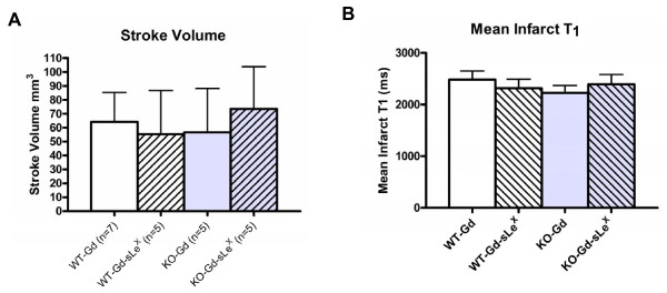
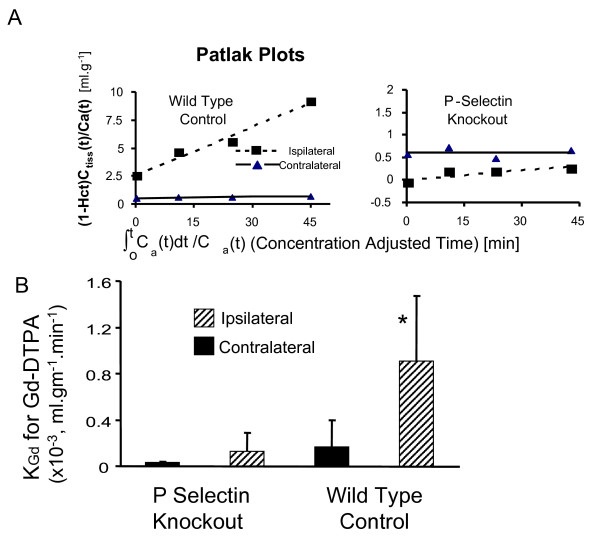
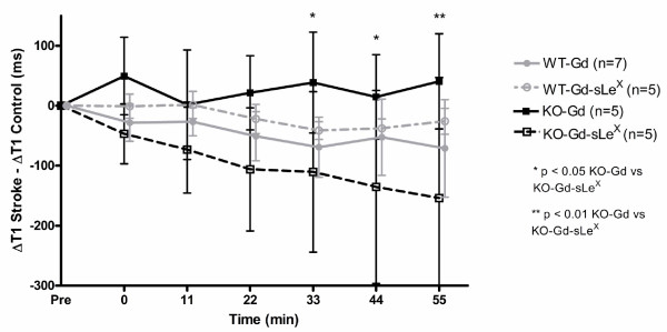
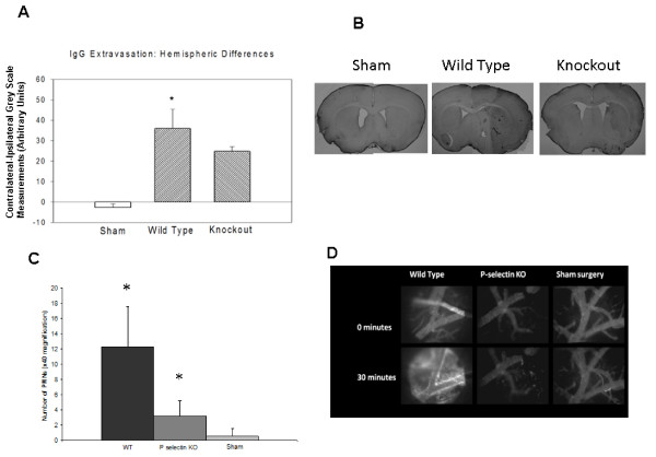
Results: MRI confirmed similar infarct sizes and T1 values at 24 hours following stroke for both WT and KO animals. However, the blood to brain transfer constant for Gd DTPA (Kgd) demonstrated greater tissue extravasation of Gd DTPA in WT animals than KO mice (P < 0.03). In the P selectin KO mice, Delta T1 stroke -Delta T1 contralateral control cortex, decreased significantly in the Gd-DTPA(sLeX) group compared to Gd-DTPA, indicative of sLeX mediated accumulation of the targeted contrast agent. Regarding BBB function, in the P-selectin KO mice compared to WT control mice, there was an attenuation in the extravasation of IgG (P < 0.001), a trend for decreased FITC extravasation and less infiltration of PMN leukocytes (P < 0.001) thereby supporting the observed increase in Kgd permeability in stroke brain of WT compared to KO mice.

Conclusion: P-selectin expression contributes to enhanced BBB dysfunction at 24 hours after transient focal cerebral ischemia.


Figures

Figure 1
Figure 1
Effects of transient cerebral ischemia on mean infarct volume and mean cerebral. T1. Twenty four hours following 30 min. of middle cerebral artery occlusion infarct volumes (mean ± SD) were similar in wild-type mice (WT) or P-selectin knockout (KO) mice administered either gadolinium-DTPA (Gd) or Gd-DTPA conjugated with sialyl Lewis X (sLex) (A). Also, shown are T1 values in the ischemic brain prior to contrast administration. (mean ± SD) (B). Values were similar in all groups (n.s.).
Figure 2
Figure 2
Effect of transient focal cerebral ischemia on the transfer constant (KGd) for Gd-DTPA into brain. Shown in the upper panel (A) are the Patlak Plots used to determine KGd. The y-axis in these plots is the ratio of the tissue concentration of contrast Ctiss(t), divided by the arterial plasma concentration at that time, i.e. Ca(t)/(1-Hct), where Hct is the arterial hematocrit. The tissue concentration was determined from measures of tissue T1 and the blood concentration was estimated from T1 in the sagittal sinus. The slope provides a measure of KGd. The lower Panel (B) shows that mean KGd values for Gd-DTPA are less in P-selectin knockout animals compared to Wildtype controls. * P < 0.03, P-selectin Knockouts compared to Wild type groups, ipsilaterally.
Figure 3
Figure 3
Magnetic resonance molecular imaging with Gd-DTPA-(sLeX). In knockout (KO) mice there was increasing effects of targeted contrast Gd-DTPA-(sLeX) on tissue T1 levels in the stroke compared to the contralateral control hemisphere not observed with non-targetted contrast or Gd-DTPA. *P < 0.05 and **P < 0.01. In wild type (WT) animals the administration of Gd-DTPA-(sLeX) or Gd-DTPA resulted in a decrease in T1 in the stroke compared to contralateral control hemisphere in both groups but there no statistical difference between groups.
Figure 4
Figure 4
IgG extravasation and polymorphonuclear (PMN) leukocyte infiltration after acute stroke. The contralateral - ipsilateral difference in IgG staining was greater in wild-type than in sham surgery or P-selectin-deficient mice (A), indicating more severe BBB injury in mice expressing P-selectin (* p < 0.05 for wild-type vs. knockout (KO) mice). Also shown are representative sections stained for IgG immunohistochemistry from sham, wild type and P selectin KO mice (B). Numbers PMN leukocytes was greater in ischemic brains of wild-type animals compared to those in P-selectin deficient animals or shams (C) (* p < 0.05, vs sham). Representative intravital microscopy images of the pial vessels obtained either immediately or 30 minutes after intravenous administration of FITC-dextran (2000 kDa) in a wild-type mouse or a P-selectin knockout mouse at 24 hours after transient middle cerebral artery occlusion or a mouse subjected to sham surgery (D), FITC extravasation is evident in the wild-type mouse expressing P-selectin but not in the P-selectin KO animal.

Similar articles

Cited by

References

    1. Huang J, Upadhyay UM, Tamargo RJ. Inflammation in stroke and focal cerebral ischemia. Surg Neurol. 2006;66:232–245. doi: 10.1016/j.surneu.2005.12.028. - DOI - PubMed
    1. Latour LL, Kang DW, Ezzeddine MA, Chalela JA, Warach S. Early blood-brain barrier disruption in human focal brain ischemia. Ann Neurol. 2004;56:468–477. doi: 10.1002/ana.20199. - DOI - PubMed
    1. Persidsky Y, Ramirez SH, Haorah J, Kanmogne GD. Blood-brain barrier: structural components and function under physiologic and pathologic conditions. J Neuroimmune Pharmacol. 2006;1:223–236. doi: 10.1007/s11481-006-9025-3. - DOI - PubMed
    1. Ishikawa M, Cooper D, Arumugam TV, Zhang JH, Nanda A, Granger DN. Platelet-leukocyte-endothelial cell interactions after middle cerebral artery occlusion and reperfusion. J Cereb Blood Flow Metab. 2004;24:907–915. doi: 10.1097/01.WCB.0000132690.96836.7F. - DOI - PubMed
    1. Ishikawa M, Cooper D, Russell J, Salter JW, Zhang JH, Nanda A, Granger DN. Molecular determinants of the prothrombogenic and inflammatory phenotype assumed by the postischemic cerebral microcirculation. Stroke. 2003;34:1777–1782. doi: 10.1161/01.STR.0000074921.17767.F2. - DOI - PubMed

Publication types

MeSH terms