. 2010 Jan 29;37(2):159-71.
doi: 10.1016/j.molcel.2009.12.023.

The Wilms' tumor suppressor protein WT1 is processed by the serine protease HtrA2/Omi

Affiliations

The Wilms' tumor suppressor protein WT1 is processed by the serine protease HtrA2/Omi

Jörg Hartkamp et al. Mol Cell. .

Abstract

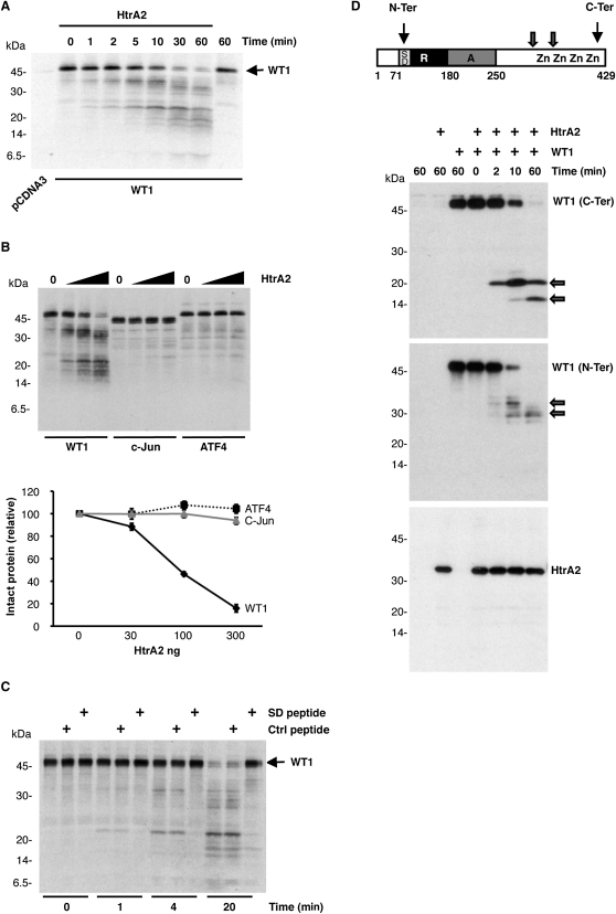
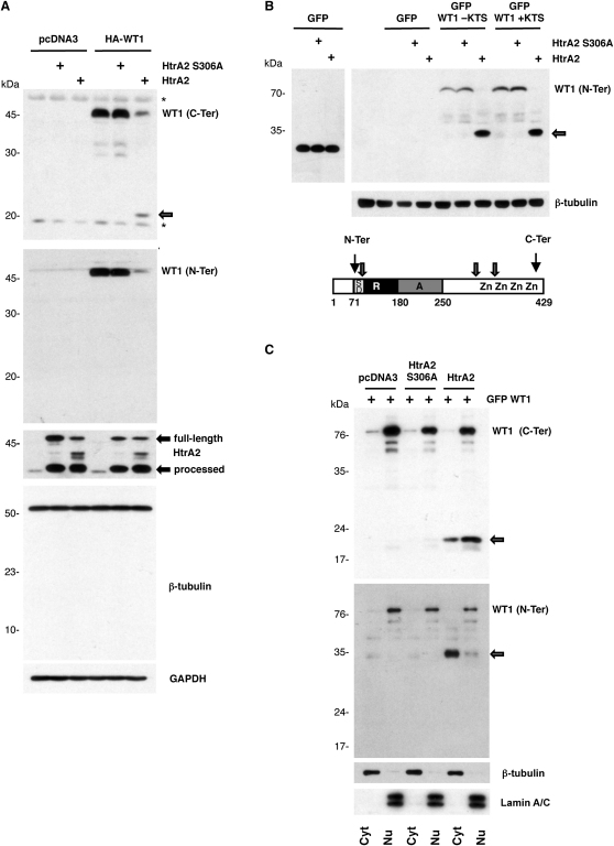
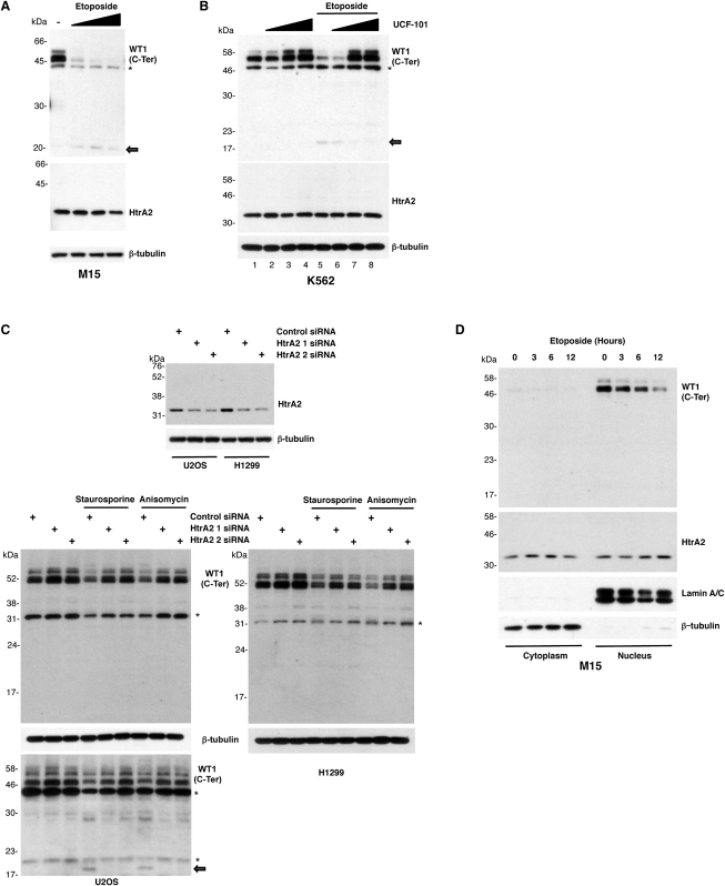
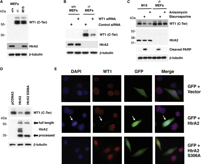
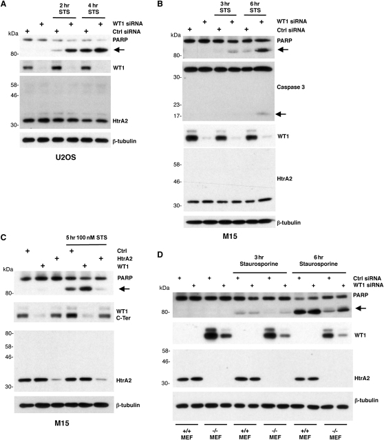
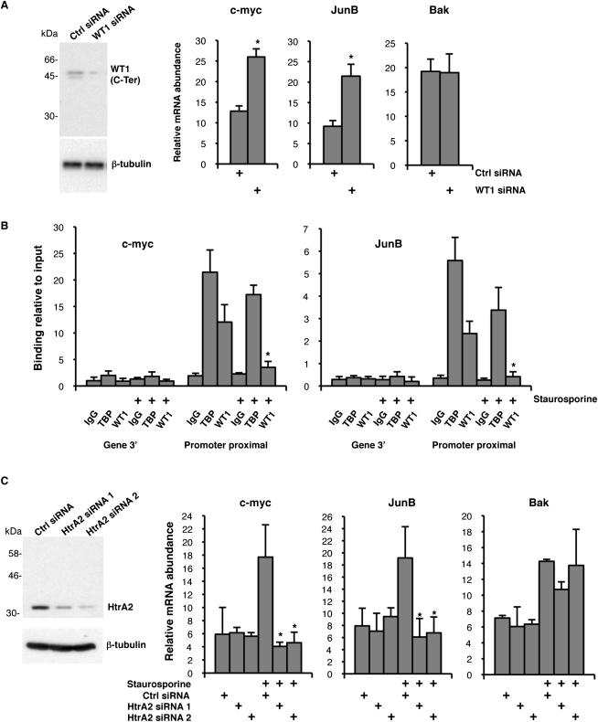
The Wilms' tumor suppressor protein WT1 functions as a transcriptional regulator of genes controlling growth, apoptosis, and differentiation. It has become clear that WT1 can act as an oncogene in many tumors, primarily through the inhibition of apoptosis. Here, we identify the serine protease HtrA2 as a WT1 binding partner and find that it cleaves WT1 at multiple sites following the treatment of cells with cytotoxic drugs. Ablation of HtrA2 activity either by chemical inhibitor or by siRNA prevents the proteolysis of WT1 under apoptotic conditions. Moreover, the apoptosis-dependent cleavage of WT1 is defective in HtrA2 knockout cells. Proteolysis of WT1 by HtrA2 causes the removal of WT1 from its binding sites at gene promoters, leading to alterations in gene regulation that enhance apoptosis. Our findings provide insights into the function of HtrA2 in the regulation of apoptosis and the oncogenic activities of WT1.


Figures

Figure 1
Figure 1
WT1 Is Processed by HtrA2 In Vitro (A) In vitro translated 35S-labeled WT1 was incubated for the indicated time with or without recombinant HtrA2 (100 ng). Samples were resolved by SDS-PAGE followed by autoradiography. (B) 35S-labeled WT1, c-Jun, and ATF4 were incubated for 30 min with increasing amounts of recombinant wild-type HtrA2 (30, 100, and 300 ng) and analyzed as described in (A). The data are presented graphically below the autoradiogram as remaining intact WT1 with increasing HtrA2 (determined by densitometry). Note that the x axis is not linear. Error bars show standard deviation from the mean (SDM). (C) 35S-labeled WT1 was incubated for the indicated time with recombinant wild-type HtrA2 (100 ng) and 1 μg of a synthetic peptide containing the WT1 suppression domain (SD, EQCLSAFTLHSFGQFTGT, residues 86–103 of WT1), a control peptide (MAAPLLHTRLPGDAC), or no peptide and was analyzed as described in (A). (D) In vitro translated WT1 was incubated for the indicated time with recombinant wild-type HtrA2 (100 ng). Samples were immunoblotted with WT1 N-Ter, C-Ter, or HtrA2 antibodies. At top, a diagram of WT1 is shown, indicating the transcriptional regulatory and zinc finger regions. The WT1 N-Ter and C-Ter epitopes and also the approximate locations of HtrA2 cleavage sites are indicated.
Figure 2
Figure 2
HtrA2 Cleaves WT1 in Cells (A) HeLa cells were cotransfected with plasmids encoding WT1 (1 μg) and vector control, along with plasmid driving expression of either wild-type or the mutant HtrA2 derivative S306A (0.5 μg). At 24 hr after transfection, whole-cell lysates were prepared and immunoblotted with the indicated antibodies. The full-length and processed forms of HtrA2 are indicated. Nonspecific bands are indicated by asterisks. (B) HeLa cells were cotransfected with plasmids encoding GFP-WT1-KTS, GFP-WT1 + KTS, or GFP (1 μg) in combination with vector control, wild-type, or mutant HtrA2 (0.5 μg) and 24 hr after transfection were analyzed as in (A). Below, a diagram of WT1 is shown, indicating the transcriptional regulatory and zinc finger regions. The epitopes recognized by the WT1 N-Ter and C-Ter antibodies and also the approximate locations of HtrA2 cleavage sites are indicated. (C) HeLa cells were transfected as in (B), and then nuclear (Nu) and cytoplasmic (Cyt) extracts were prepared. The samples were immunoblotted with the antibodies indicated. The GFP-WT1 N-terminal and C-terminal proteolytic fragments are indicated.
Figure 3
Figure 3
HtrA2 Is Required for the Stimulation of WT1 Cleavage following Treatment with Cytotoxic Drugs (A) Mouse embryonic M15 cells were treated for 20 hr with 10, 30, and 100 μM Etoposide and analyzed by immunoblotting with WT1 antibodies (C-ter). The 20 kDa cleavage product is indicated by an arrow. Nonspecific bands are indicated by asterisks. (B) Human K562 cells were incubated with the HtrA2-specific inhibitor UCF-101 (15, 30, and 45 μM) for 30 min before stimulation with 100 μM Etoposide for 24 hr and were analyzed as in (A). Nonspecific bands are indicated by asterisks. (C) U2OS (left) or H1299 cells (right) were transfected with control siRNA or with two different HtrA2-specific siRNAs. HtrA2 knockdown was confirmed by immunoblotting. At 60 hr after transfection, the cells were stimulated with 0.5 μM Staurosporine or 5 ng/ml Anisomycin for 8 hr and analyzed by immunoblot with the antibodies indicated. The lower immunoblot for the U2OS cells used a different anti-WT1 (C-Ter) antibody that is more sensitive, and the 20 kDa C-terminal proteolytic fragment is indicated. (D) Untreated M15 cells or M15 cells that had been treated with 30 μM etoposide for the time points indicated were fractionated to produce nuclear (Nu) and cytoplasmic (Cyt) fractions. The samples were immunoblotted with the antibodies indicated.
Figure 4
Figure 4
Analysis of WT1 in HtrA2 Null MEFs (A) Whole-cell lysates were prepared from HtrA2+/+ or HtrA2−/− mouse embryonic fibroblasts (MEFs) and compared with mouse M15 cells by immunoblotting. (B) HtrA2+/+ or HtrA2−/− MEFs were transfected with control siRNA or with WT1-specific siRNA, and 30 hr later, whole-cell lysates were prepared and immunoblotted with the antibodies indicated. (C) M15 cells or HtrA2−/− MEFs were stimulated for 8 hr with 0.5 μM Staurosporine or 5 ng/ml Anisomycin, and whole-cell lysates were prepared and immunoblotted with the antibodies indicated. (D) HtrA2−/− MEFS were transfected with control vector (pCDNA3), vector driving expression of wild-type HtrA2, or the HtrA2 mutant derivative S306A. Whole-cell lysates were immunoblotted with the antibodies indicated. The full-length and processed forms of HtrA2 are indicated. (E) HtrA2−/− MEFS were cotransfected with a vector driving expression of GFP along with either control vector (pCDNA3) and vector driving expression of wild-type HtrA2 or the HtrA2 mutant derivative S306A. The cells were analyzed by immunofluorescence with anti-WT1 antibodies (red) and overlayed with the GFP signal.
Figure 5
Figure 5
WT1 Is an Inhibitor of Apoptosis (A) U2OS cells were transfected with a control siRNA or siRNA that targets WT1. At 24 hr later, the cells were treated with 100 nM Staurosporine for the indicated times. Whole-cell extracts were prepared and immunoblotted with the antibodies indicated. (B) M15 cells were analyzed as in (A) except that the siRNAs were mouse specific. The samples were also immunoblotted with caspase 3 antibody. (C) M15 cells were transfected with control or HtrA2 siRNA, and 24 hr later, were split 1:4. A further 24 hr later, the cells were transfected with control or WT1 siRNA, and then 24 hr later, they were treated with 100 nM Staurosporine for 5 hr and harvested, and whole-cell extracts were subjected to immunoblotting with the antibodies indicated. (D) MEFs were transfected with control or WT1-specific siRNA, and 24 hr later, they were treated with 100 nM Staurosporine for the indicated time, and then whole-cell lysates were prepared and analyzed by immunoblotting with the antibodies indicated.
Figure 6
Figure 6
Degradation of WT1 by HtrA2 Releases the c-myc and JunB Genes from Transcriptional Repression by WT1 (A) U2OS cells were transfected as in Figure 5A and either used to prepare whole-cell extracts for immunoblotting with the indicated antibodies or to prepare total RNA and then cDNA. The expression of c-myc, JunB, and Bak are shown graphically relative to GAPDH expression. Error bars show standard deviation from the mean (SDM). p < 0.05 in Student's t test. (B) U2OS cells were subject to ChIP with the antibodies indicated before and after treatment with 0.5 μM Staurosprine for 12 hr. Binding of WT1 and TBP to the c-myc (left graph) and JunB (right graph) promoters or the 3′ gene regions was quantitated by PCR and expressed as percent enrichment of the input chromatin. Error bars show SDM. p < 0.05 in Student's t test. (C) U2OS cells were treated with 200 nM Staurosporine following transfection with either control siRNA or two different HtrA2-specific siRNAs. Total RNA was prepared and quantitative PCR was performed to analyze c-myc, JunB, and Bak expression relative to GAPDH. Error bars show SDM. p < 0.05 in Student's t test. An immunoblot of HtrA2 is shown at left, confirming robust knockdown of HtrA2 by the siRNAs.

Comment in

Similar articles

Cited by

References

    1. Ariyaratana S., Loeb D.M. The role of the Wilms tumour gene (WT1) in normal and malignant haematopoiesis. Expert Rev. Mol. Med. 2007;9:1–17. - PubMed
    1. Carpenter B., Hill K.J., Charalambous M., Wagner K.J., Lahiri D., James D.I., Andersen J.S., Schumacher V., Royer-Pokora B., Mann M. BASP1 is a transcriptional cosuppressor for the Wilms' tumor suppressor protein WT1. Mol. Cell. Biol. 2004;24:537–549. - PMC - PubMed
    1. Cilenti L., Lee Y., Hess S., Srinivasula S., Park K.M., Junqueira D., Davis H., Bonventre J.V., Alnemri E.S., Zervos A.S. Characterization of a novel and specific inhibitor for the pro-apoptotic protease Omi/HtrA2. J. Biol. Chem. 2003;278:11489–11494. - PubMed
    1. Discenza M.T., Pelletier J. Insights into the physiological role of WT1 from studies of genetically modified mice. Physiol. Genomics. 2004;16:287–300. - PubMed
    1. Dutton J.R., Lahiri D., Ward A. Different isoforms of the Wilms' tumour protein WT1 have distinct patterns of distribution and trafficking within the nucleus. Cell Prolif. 2006;39:519–535. - PMC - PubMed

Publication types

LinkOut - more resources