

Comparative Study
doi: 10.1016/j.leukres.2009.12.013.
Epub 2010 Jan 31.
Comparative analysis of MVA-CD40L and MVA-TRICOM vectors for enhancing the immunogenicity of chronic lymphocytic leukemia (CLL) cells
Affiliations
- PMID: 20122733
- PMCID: PMC2891581
- DOI: 10.1016/j.leukres.2009.12.013
Item in Clipboard
Comparative Study
Comparative analysis of MVA-CD40L and MVA-TRICOM vectors for enhancing the immunogenicity of chronic lymphocytic leukemia (CLL) cells
Leuk Res.
2010 Oct.
Abstract
Adenoviral transduction with CD40L and poxviral transduction with B7-1, ICAM-1, and LFA-3 (TRICOM) have been used to enhance the antigen-presenting capacity of chronic lymphocytic leukemia (CLL) cells. This study compares the same vector (modified vaccinia virus strain Ankara (MVA)) encoding CD40L or TRICOM for its ability to enhance the immunogenicity of CLL cells. CLL cells from some patients showed differential responses to each vector in terms of induction of autologous T-cell responses. This study supports the rationale for the use of CLL cells modified ex vivo with pre-specified recombinant MVA vectors as a whole tumor-cell vaccine for immunotherapy in CLL patients.
Published by Elsevier Ltd.
Figures
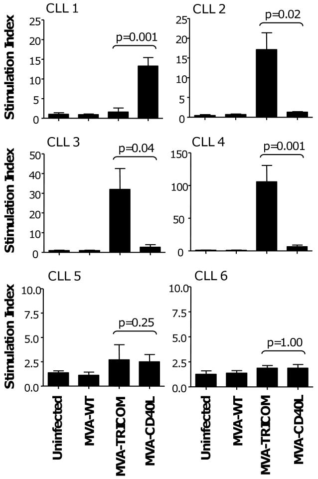
CLL cells from 4 patients were infected with MVA-WT, MVA-TRICOM, or MVA-CD40L. Following 24 hours of infection, CLL cells were washed, irradiated, and cocultured with allogeneic CD3+ T cells from a healthy donor at a ratio of effector to stimulator cells of 10:1. Error bars indicate the standard deviation of replicate measurements. Note that, due to variability in responses, different scales were used on the graphs.
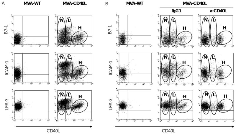
(A) CLL cells were infected with MVA-WT or MVA-CD40L. Following 24 hours of infection, CLL cells were analyzed by flow cytometry for expression of B7-1, ICAM-1, and LFA-3 by CD40L-expressing and non-expressing populations. The plots shown were gated on CD19+ B cells. After infection with MVA-CD40L, 3 populations of CLL cells that expressed negative, low, or high levels of CD40L were observed; these are designated as “negative” (N), “low MFI” (L), and “high MFI” (H) populations on the plots shown. (B) CLL cells were treated with blocking antibody (anti-CD40L or isotype-control mouse IgG1) before and during infection with MVA-CD40L.
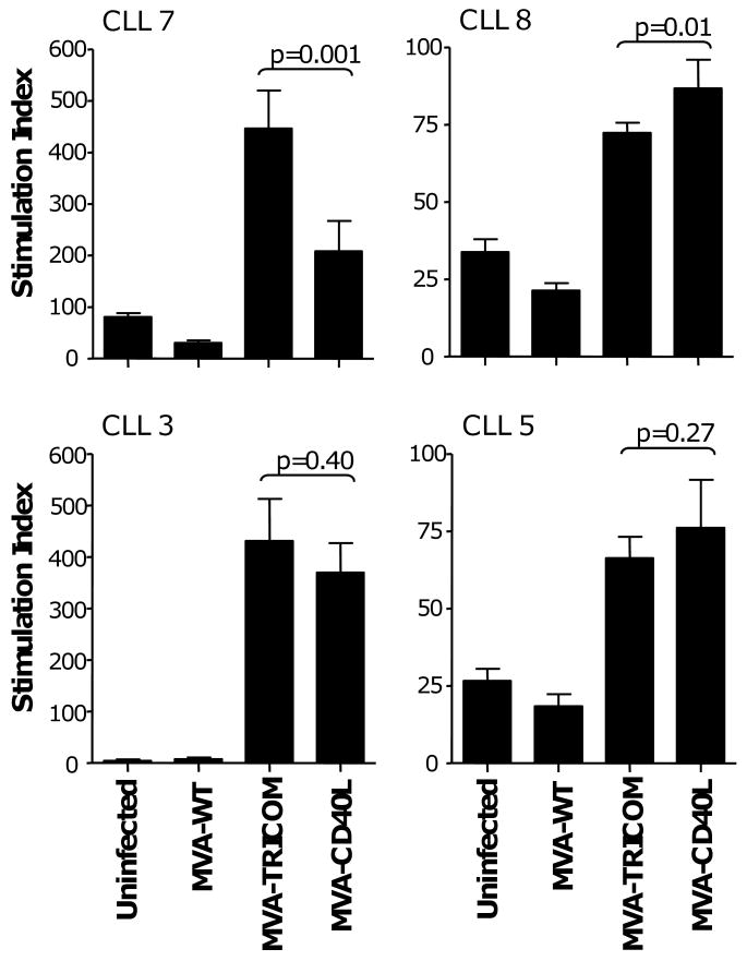
CLL cells from 6 patients were infected with MVA-WT, MVA-TRICOM, or MVA-CD40L. Following 24 hours of infection, CLL cells were washed, irradiated, and cocultured with autologous CD3+ T cells at a ratio of effector to stimulator cells of 2.5:1. Error bars indicate the standard deviation of replicate measurements. Note that, due to variability in responses, different scales were used on the graphs. Costimulatory molecule expression by the CLL cells following infection is shown in Table 1.
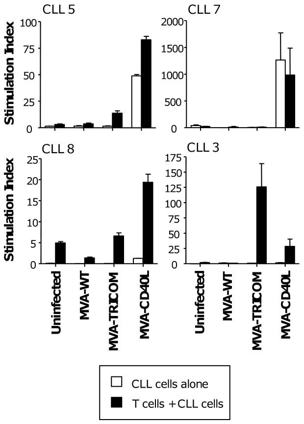
CLL cells from 4 patients were infected with MVA-WT, MVA-TRICOM, or MVA-CD40L. Following 24 hours of infection, CLL cells were washed and, without irradiation, cocultured with autologous CD3+ T cells at a ratio of effector to stimulator cells of 2.5:1. Proliferation by the CLL cells alone is indicated by white bars; proliferation by T cells and CLL cells together is indicated by black bars. Error bars indicate the standard deviation of replicate measurements. Note that, due to variability in responses, different scales were used on the graphs.
References
-
- Wierda WG, Kipps TJ. Chronic lymphocytic leukemia. Curr Opin Hematol. 1999;6:253–61. - PubMed
-
- Tsimberidou AM, Keating MJ. Treatment of fludarabine-refractory chronic lymphocytic leukemia. Cancer. 2009;115:2824–36. - PubMed
-
- Pleyer L, Egle A, Hartmann TN, Greil R. Molecular and cellular mechanisms of CLL: novel therapeutic approaches. Nat Rev Clin Oncol. 2009;6:405–18. Epub 2009 Jun 2. - PubMed
-
- Buhmann R, Nolte A, Westhaus D, Emmerich B, Hallek M. CD40-activated B-cell chronic lymphocytic leukemia cells for tumor immunotherapy: stimulation of allogeneic versus autologous T cells generates different types of effector cells. Blood. 1999;93:1992–2002. - PubMed
-
- Rezvany MR, Jeddi-Tehrani M, Rabbani H, Lewin N, Avila-Carino J, Osterborg A, et al. Autologous T lymphocytes may specifically recognize leukaemic B cells in patients with chronic lymphocytic leukaemia. Br J Haematol. 2000;111:608–17. - PubMed
Publication types
MeSH terms
Substances
Grants and funding
LinkOut - more resources
Full Text Sources
Other Literature Sources
Miscellaneous
