

Factors associated with improvement and worsening of visual acuity 2 years after focal/grid photocoagulation for diabetic macular edema
- PMID: 20122739
- PMCID: PMC2864322
- DOI: 10.1016/j.ophtha.2009.10.002
Factors associated with improvement and worsening of visual acuity 2 years after focal/grid photocoagulation for diabetic macular edema
Abstract
Purpose: To identify factors associated with the visual acuity outcome after focal/grid photocoagulation for diabetic macular edema (DME) among eyes randomized to the focal/grid photocoagulation treatment group within the Diabetic Retinopathy Clinical Research Network (DRCR.net) trial comparing triamcinolone with focal/grid laser.
Design: Multicenter, randomized, clinical trial.
Participants: Three hundred thirty eyes with DME assigned to the focal/grid photocoagulation group, visual acuity 20/40 to 20/320, and optical coherence tomography (OCT) central subfield thickness > or =250 microns.
Methods: Eyes were treated with a protocol-defined photocoagulation technique, which was repeated at 4-month intervals for persistent or recurrent edema. Separate logistic regression models were used to evaluate the associations of demographic, clinical, OCT, and fundus photographic variables with visual acuity improvement or worsening of > or =10 letters from baseline to 2 years. The association of the initial visual acuity outcome after treatment with the subsequent visual acuity course also was evaluated.
Main outcome measures: Visual acuity measured with the electronic Early Treatment Diabetic Retinopathy Study method.
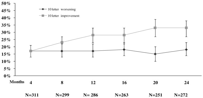
Results: Worse baseline visual acuity was the only factor found to be associated with more frequent visual acuity improvement (P<0.001), and both greater baseline OCT-measured retinal volume (P = 0.001) and better baseline visual acuity (P = 0.009) were found to be associated with more frequent visual acuity worsening. Visual acuity outcomes were similar in eyes with and without prior macular or panretinal photocoagulation. The initial visual acuity outcome at 4 months was not generally predictive of the subsequent course. Many eyes that worsened > or =10 letters from baseline to 4 months subsequently improved, and many eyes that initially improved, subsequently worsened.
Conclusions: At this time, focal/grid photocoagulation remains the standard management for DME and these results do not alter this paradigm.
Copyright 2010 American Academy of Ophthalmology. Published by Elsevier Inc. All rights reserved.
Figures
References
-
- Diabetes Control and Complications Trial Research Group. The effect of intensive diabetes treatment on the progression of diabetic retinopathy in insulin-dependent diabetes mellitus: the Diabetes Control and Complications Trial. Arch Ophthalmol. 1995;113:36–51. - PubMed
-
- Klein R, Klein BE. Vision disorders in diabetes. In: National Diabetes Data Group, editor. Diabetes in America. 2nd. Bethesda, MD: National Institute of Diabetes and Digestive and Kidney Diseases; 1995. [August 21, 2009]. pp. 293–338. NIH publ. no. 95–1468 Available at: http://diabetes.niddk.nih.gov/dm/pubs/america/pdf/chapter14.pdf.
-
- Klein R, Klein BE, Moss SE, et al. The Wisconsin Epidemiologic Study of Diabetic Retinopathy. IV. Diabetic macular edema. Ophthalmology. 1984;91:1464–74. - PubMed
-
- Moss SE, Klein R, Klein BE. The 14-year incidence of visual loss in a diabetic population. Ophthalmology. 1998;105:998–1003. - PubMed
Publication types
MeSH terms
Substances
Grants and funding
LinkOut - more resources
Full Text Sources
Medical
