Review
. 2010 Apr;13(2):124-32.
doi: 10.1016/j.mib.2009.12.014. Epub 2010 Feb 1.

Bacterial chemoreceptors: providing enhanced features to two-component signaling

Affiliations
Review

Bacterial chemoreceptors: providing enhanced features to two-component signaling

Gerald L Hazelbauer et al. Curr Opin Microbiol. 2010 Apr.

Abstract

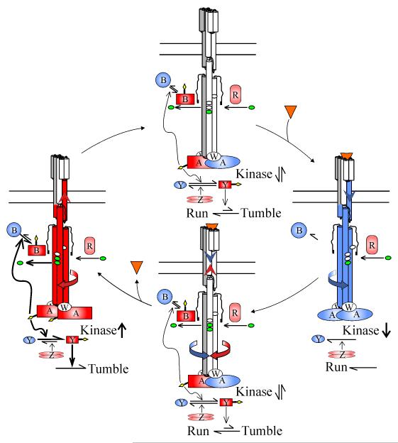
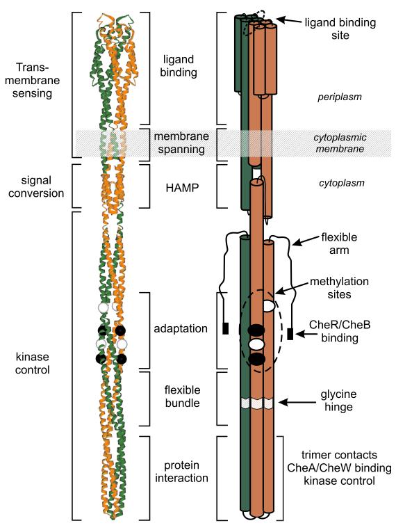
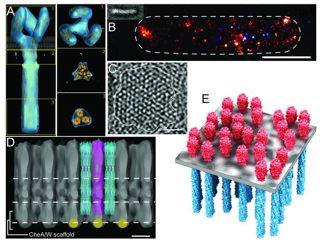
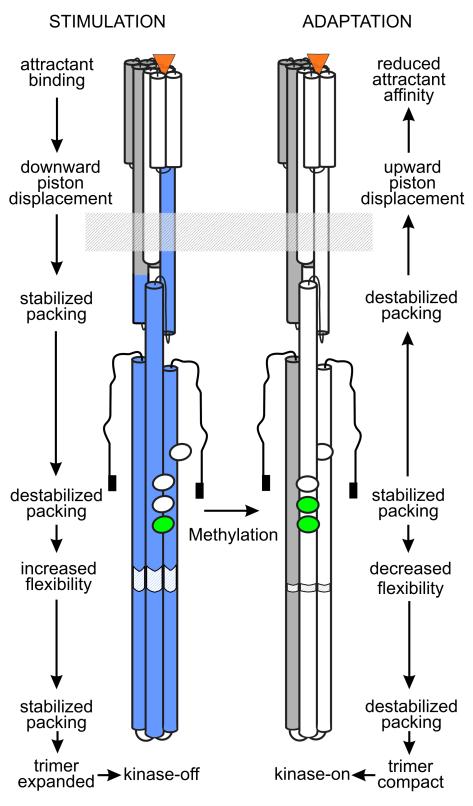
Bacteria perform chemotaxis utilizing core two-component signaling systems to which have been added enhanced features of signal amplification, sensory adaptation, molecular memory and high sensitivity over a wide dynamic range. Chemoreceptors are central to the enhancements. These transmembrane homodimers associate in trimers and in clusters of signaling complexes containing from a few to thousands of receptors. Receptor homodimers couple ligand occupancy and adaptational modification to transmembrane signaling. Trimers activate and control the histidine kinase. Clusters enable signal amplification, high sensitivity and adaptational assistance. Homodimer signaling initiates with helical piston sliding that is converted to modulation of competing packing modes of adjacent segments of an extended helical coiled coil. In trimers, signaling and coupling may involve switching between compact and expanded forms.


Figures

Fig. 1
Fig. 1. The strategy of the chemotaxis sensory system of E. coli
The figure shows the processes of stimulation and adaptation for a positive stimulus (increase in attractant concentration; right and bottom panel) and a negative stimulus (decrease in attractant concentration; left and top panel) using cartoons of the signaling complex representing the two receptor conformations (left = “kinase-on”, right = “kinase-off”). See text for discussion of the equilibrium between these two conformations. For clarity, only one of the three receptor dimers in the signaling complex is shown. Che proteins are labeled by their respective letters. Methyl-accepting sites are marked by ovals on the chemoreceptor, methyl groups are green and phosphoryl groups are yellow diamonds. Red symbolizes activated or activating forms, blue inactive or inactivating forms and pink constitutive activity. Top panel: Methyltransferase CheR adds methyl groups to the receptor and methylesterase CheB, activated by CheA-mediated phosphorylation, removes them. Phospho-CheY (CheY-P) binds to the flagellar rotary motor, switching its default state of counter-clockwise to clockwise and thus motility from straight-line runs to swimming-direction-reorienting tumbles. A balance between creation of CheY-P by CheA and destruction by phosphatase CheZ results in a steady state concentration of CheY-P that produces tumble episodes every few seconds, causing the cell to reorient and thus trace a three-dimensional random walk. Right panel: Attractant (orange triangles) binding to a periplasmic site generates a piston movement in the transmembrane sensing module (blue helix and arrowhead) which induces a cascade of alternating stabilizing and destabilizing changes in the helical packing of the signal conversion and kinase control modules (blue helixes and curving arrow) to shift the receptor conformational equilibrium toward the kinase-off, methylation-on, demethylation-off receptor conformation. Kinase inhibition reduces the cellular content of short-lived CheY-P and CheB-P, reducing the probability of tumbles and demethylation, respectively. Bottom panel: Altered receptor propensities for methylation and demethylation plus reduction of CheB-P result in an increase in receptor methylation that counteracts (opposing red arrows) the effects of ligand binding and thus returns the conformational equilibrium, propensities for adaptational modification, kinase activity, CheY-P, CheB-P and motile behavior to their null states. Left panel: Loss of ligand eliminates the changes generated by receptor occupancy but the compensatory effects of increased methylation are still present (red arrows), driving the receptor toward a kinase-on, methylation-off, demethylation-on conformation (red arrows and helices) that results in heightened kinase activity and thus higher levels of CheY-P and CheB-P, generating increased tumbling and demethylation, respectively. Effects on receptor propensities for modification and methylesters activity result in receptor methylation dropping to the original value in the absence of stimulation, thus reestablishing the adapted state (top panel).
Fig. 2
Fig. 2. The chemoreceptor dimer
A ribbon diagram (left) and a cartoon (right) of a chemoreceptor dimer illustrate structural and functional features. Modules are identified to the left of the ribbon diagram, functions or motifs are indicated between the ribbon diagram and cartoon, and specific features noted to the right of the cartoon. See text for details. The figure is based on Fig. 1, Box 3 of [1].
Fig. 3
Fig. 3. Higher order interactions of chemoreceptor dimers
A. Trimer structure. The panels show the “compact” conformation detected by cryo-electron tomography of E. coli chemoreceptors in a crystalline array, fit with three receptor dimers [20••]. The left-hand panel is a view perpendicular to the long axis of the receptor with the position of the cytoplasmic membrane indicated. The three right-hand panels are views parallel to the membrane at the three positions indicated in the left-hand image. The panels are from Fig. 4 of ref. [20••] with permission. B. Distribution of signaling complexes in an E. coli cell imaged by photoactivated localization microscopy (PALM). Chemotactically functional CheW fused to a photoactivatable fluorescent protein is visualized in a fixed cell by a procedure that can detect each individual molecule in a field of view. The image is Fig. 3D of ref. [31••] with permission. C. A tomographic slice of a face-on view of a chemoreceptor patch in C. cresentus showing partially ordered hexagonal packing. The image is Fig. 4A of ref. [11•] with permission. D. An edge-on view of the densities of an averaged packing arrangement of trimeric receptors in a C. crescentus signaling complex patch. Although beyond the scope of this review, it is tantalizing to note that the averaged densities imply that CheA and CheW associate with every second receptor trimer. The image is Fig. 3B of ref. [11•] with permission. E. Model derived from tomographic analysis of the organization of chemoreceptor trimers of dimers in patches of C. cresentus signaling complexes. The image is Fig. 4C of ref. [11•] with permission.
Fig. 4
Fig. 4. Chemoreceptor signaling
Cartoons of a stimulated and adapted receptor dimer illustrate our understanding, explained in the text, of the conformational changes that couple stimulation by attractant binding to kinase inhibition (left-hand image and labels) and adaptation by methylation to re-establish pre-stimulation kinase activity (right-hand image and labels) . See Fig. 2 for labels of the receptor features shown in the cartoon and Fig. 1 for the symbolism of the colors. The figure is based on Fig. 3 of [1].

Similar articles

Cited by

References

    1. Hazelbauer GL, Falke JJ, Parkinson JS. Bacterial chemoreceptors: high-performance signaling in networked arrays. Trends Biochem Sci. 2008;33:9–19. - PMC - PubMed
    1. Szurmant H, Ordal GW. Diversity in chemotaxis mechanisms among the bacteria and archaea. Microbiol Mol Biol Rev. 2004;68:301–319. - PMC - PubMed
    1. Wadhams GH, Armitage JP. Making sense of it all: bacterial chemotaxis. Nat Rev Mol Cell Biol. 2004;5:1024–1037. - PubMed
    1. Rao CV, Glekas GD, Ordal GW. The three adaptation systems of Bacillus subtilis chemotaxis. Trends Microbiol. 2008;16:480–487. - PMC - PubMed
    1. Vladimirov N, Sourjik V. Chemotaxis: how bacteria use memory. Biol Chem. 2009;390:1097–1104. - PubMed

Publication types

MeSH terms