. 2010 Mar 19;397(1):161-78.
doi: 10.1016/j.jmb.2010.01.059. Epub 2010 Feb 1.

Multiple nucleotide preferences determine cleavage-site recognition by the HIV-1 and M-MuLV RNases H

Affiliations

Multiple nucleotide preferences determine cleavage-site recognition by the HIV-1 and M-MuLV RNases H

Sharon J Schultz et al. J Mol Biol. .

Abstract

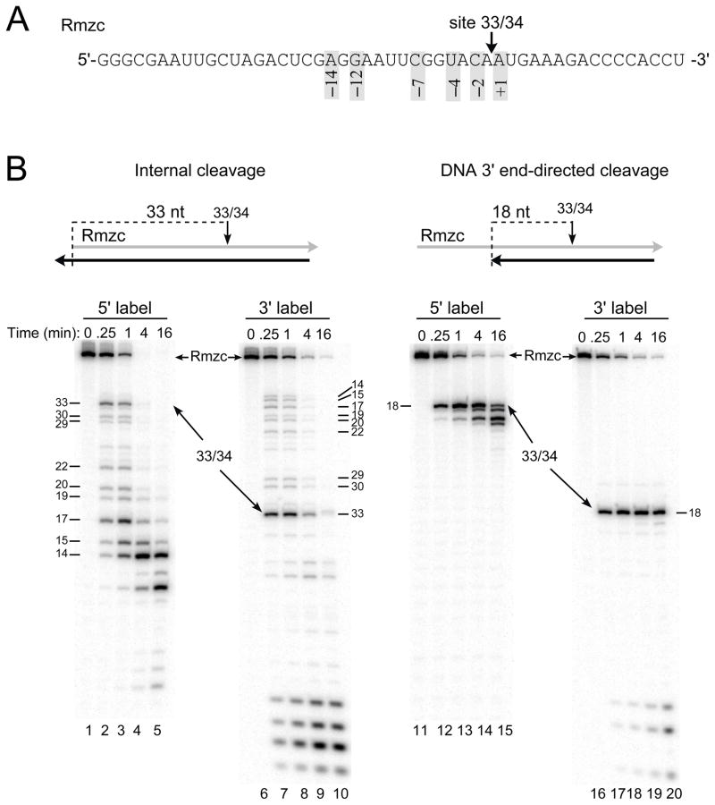
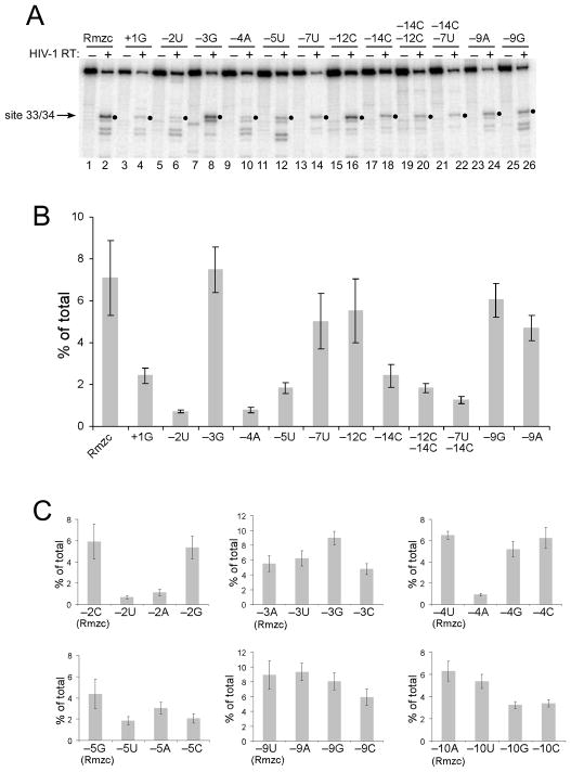
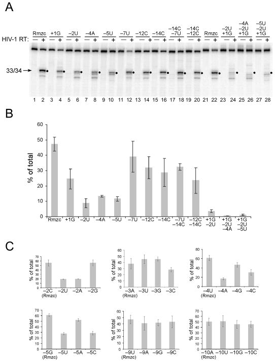
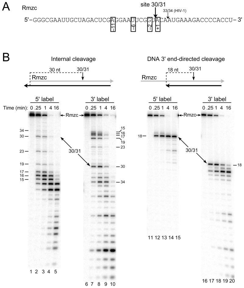
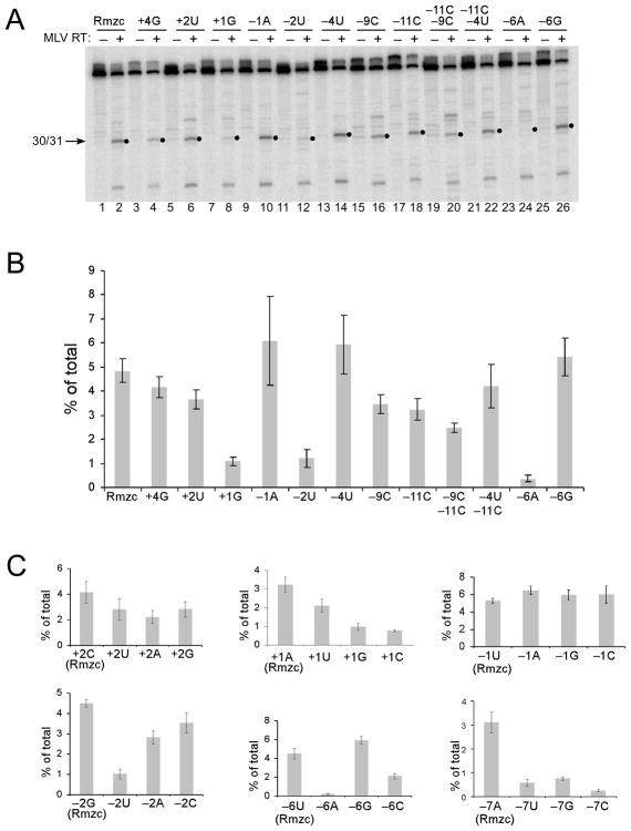
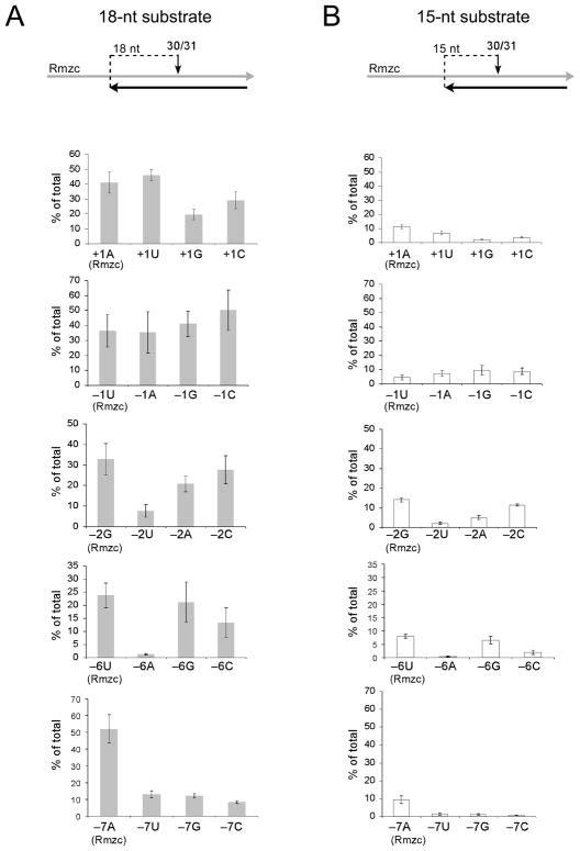
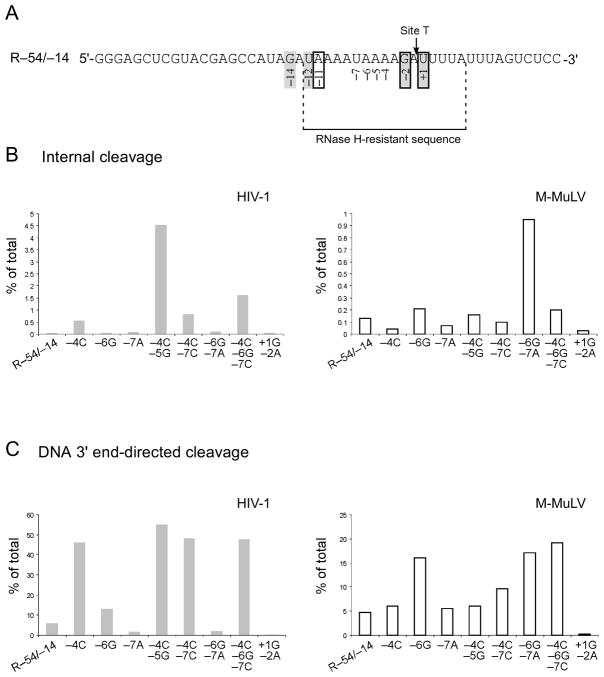
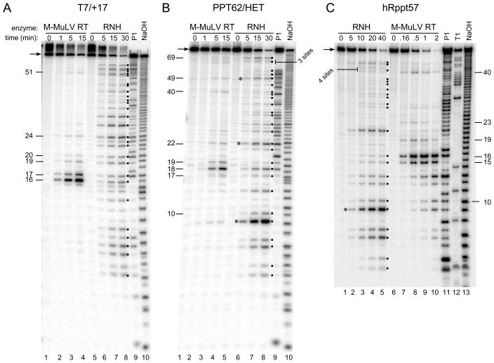
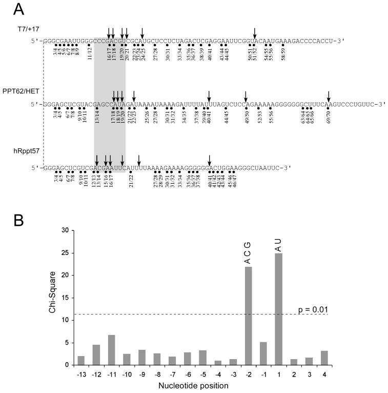
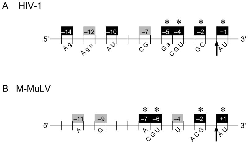
The RNase H activity of reverse transcriptase is required during retroviral replication and represents a potential target in antiviral drug therapies. Sequence features flanking a cleavage site influence the three types of retroviral RNase H activity: internal, DNA 3'-end-directed, and RNA 5'-end-directed. Using the reverse transcriptases of HIV-1 (human immunodeficiency virus type 1) and Moloney murine leukemia virus (M-MuLV), we evaluated how individual base preferences at a cleavage site direct retroviral RNase H specificity. Strong test cleavage sites (designated as between nucleotide positions -1 and +1) for the HIV-1 and M-MuLV enzymes were introduced into model hybrid substrates designed to assay internal or DNA 3'-end-directed cleavage, and base substitutions were tested at specific nucleotide positions. For internal cleavage, positions +1, -2, -4, -5, -10, and -14 for HIV-1 and positions +1, -2, -6, and -7 for M-MuLV significantly affected RNase H cleavage efficiency, while positions -7 and -12 for HIV-1 and positions -4, -9, and -11 for M-MuLV had more modest effects. DNA 3'-end-directed cleavage was influenced substantially by positions +1, -2, -4, and -5 for HIV-1 and positions +1, -2, -6, and -7 for M-MuLV. Cleavage-site distance from the recessed end did not affect sequence preferences for M-MuLV reverse transcriptase. Based on the identified sequence preferences, a cleavage site recognized by both HIV-1 and M-MuLV enzymes was introduced into a sequence that was otherwise resistant to RNase H. The isolated RNase H domain of M-MuLV reverse transcriptase retained sequence preferences at positions +1 and -2 despite prolific cleavage in the absence of the polymerase domain. The sequence preferences of retroviral RNase H likely reflect structural features in the substrate that favor cleavage and represent a novel specificity determinant to consider in drug design.


Figures

Fig. 1
Fig. 1
Model substrates to analyze internal and DNA 3′ end-directed cleavage by HIV-1 reverse transcriptase. A. The sequence of 49-mer RNA Rmzc is shown from 5′ to 3′ and site 33/34, the strong cleavage site recognized by HIV-1 reverse transcriptase, is indicated with an arrow. For site 33/34, the positions of preferred nucleotides as determined from an earlier statistical analysis are indicated by the numbers in shaded boxes. B. Cleavage of site 33/34 in Rmzc hybrids designed to test internal (left panel) or DNA 3′ end-directed (right panel) cleavage. Above each panel is a schematic of the hybrid substrate containing Rmzc (gray) and a DNA strand (black). HIV-1 reverse transcriptase was incubated with hybrids containing 5′ end-labeled (5′ label; lanes 1–5, 11–15) or 3′ end-labeled Rmzc (3′ label; lanes 6–10, 16–20) in time course assays. Samples were either left untreated (0 min) or removed at 0.25, 1, 4, or 16 min, analyzed in denaturing 20% acrylamide gels, and visualized using a PhosphorImager. The positions of Rmzc and the products resulting from cleavage at site 33/34 are indicated with arrows, and the sizes of cleavage products are indicated in nucleotides.
Fig. 2
Fig. 2
Effects of nucleotide substitutions on internal RNase H cleavage by HIV-1 reverse transcriptase. A. 5′ end-labeled Rmzc or Rmzc RNAs with the indicated substitutions relative to site 33/34 were used to generate internal cleavage substrates as shown in Fig. 1B. The position(s) and change(s) introduced into each substrate relative to site 33/34 are indicated above each lane by the position number and base. These substrates were incubated with HIV-1 reverse transcriptase in time course experiments and the 1 min time points (even numbered lanes) or the untreated substrates (odd numbered lanes) were analyzed as described in Fig. 1B. The relevant portion of the resulting gel image is shown and filled circles indicate the position of the 33-mer product resulting from cleavage at site 33/34. B. Bar graph depicting the total amount of 33-mer product (% of total) generated by internal cleavage at site 33/34 using 1 min time point data from 5 separate experiments with the substrates described in A. C. Bar graphs depicting the total amount of 33-mer product (% of total) generated by internal cleavage at site 33/34 using substrates comparing cleavage of Rmzc (leftmost bar in each graph) with all three nucleotide substitutions at the indicated positions. Data from 1 min time points generated in 5 experiments are shown for all positions. For B and C, error bars represent plus and minus one standard deviation.
Fig. 3
Fig. 3
Effects of nucleotide substitutions on DNA 3′ end-directed RNase H cleavage by HIV-1 reverse transcriptase. A. DNA 3′ end-directed cleavage substrates as shown in Fig. 1B were made using 5′ end-labeled Rmzc or Rmzc RNAs with the indicated nucleotide substitutions, and the resulting substrates were analyzed as described in Fig. 2A. B. A bar graph depicting the total amount of 33-mer product (% of total) generated by DNA 3′ end-directed cleavage at site 33/34 from 1 min time points generated in 5 separate experiments using the substrates shown in A. C. Bar graphs depicting the total amount of 33-mer product (% of total) generated by DNA 3′ end-directed cleavage at site 33/34 using substrates comparing cleavage of Rmzc (left) with substrates containing the three other nucleotides at the indicated positions. Data represent the 1 min time points generated in 5 experiments (positions −2, −9, −10) or 4 experiments (positions −3, −4, −5).
Fig. 4
Fig. 4
Model substrates to analyze internal and DNA 3′ end-directed cleavage by M-MuLV reverse transcriptase. A. The sequence of Rmzc is shown from 5′ to 3′ and site 30/31, the strong cleavage site recognized by M-MuLV reverse transcriptase, is indicated just upstream of site 33/34 (used for HIV-1 RNase H assays; see Fig. 1). The positions of preferred nucleotides as determined previously are indicated by the numbers in open boxes. B. Cleavage of site 30/31 in Rmzc hybrids designed to test internal (left panel) or DNA 3′ end-directed (right panel) cleavage. The hybrid substrates are shown above each panel and configured as described in Fig. 1B. Hybrids containing Rmzc labeled at the 5′ (lanes 1–5, 11–15) or 3′ (lanes 6–10, 16–20) ends were incubated with M-MuLV reverse transcriptase in time course assays, and samples were analyzed as described in Fig. 1B. The positions of Rmzc and the 30-mer product resulting from cleavage at site 30/31 are indicated with arrows and the sizes of cleavage products are indicated in nucleotides.
Fig. 5
Fig. 5
Effects of nucleotide substitutions on internal RNase H cleavage by M-MuLV reverse transcriptase. A. 5′ end-labeled Rmzc or Rmzc RNAs with the indicated substitutions relative to site 30/31 were used to generate internal cleavage substrates as described in Fig. 2. The position(s) and change(s) introduced into each substrate relative to site 30/31 are indicated above each lane by the position number and base. These substrates were incubated with M-MuLV reverse transcriptase in time course experiments and the 0.25 min time points (even numbered lanes) or the untreated substrates (odd numbered lanes) were analyzed. Filled circles indicate the position of the 30-mer product resulting from cleavage at the 30/31 site. B. A bar graph depicting the amount of 30-mer product (% of total) generated by internal cleavage at site 30/31 from 0.25 min time points generated in 5 separate experiments using the substrates described in A. C. Bar graphs depicting the total amount of 30-mer product (% of total) generated by internal cleavage at site 30/31 using substrates comparing cleavage of Rmzc (left) with substrates containing the indicated substitutions. Data represent the 0.25 min time points generated in 5 experiments.
Fig. 6
Fig. 6
Effects of nucleotide substitutions on DNA 3′ end-directed RNase H cleavage by M-MuLV reverse transcriptase. A. DNA 3′ end-directed cleavage substrates that positioned site 30/31 at 18 nucleotides from the recessed DNA 3′ end (18-nt substrate) were made using Rmzc or Rmzc RNAs with the indicated nucleotide substitutions, and the resulting substrates were analyzed as described in Fig. 3. The bar graphs depict the amount of 30-mer product (% of total) generated by DNA 3′ end-directed cleavage at site 30/31 as described in Fig. 5C. Data represent the 0.25 min time points generated in 5 experiments (positions −1, −6, −7), 4 experiments (position +1), or 3 experiments (positions −2). B. DNA 3′ end-directed cleavage substrates that positioned site 30/31 at 15 nucleotides from the recessed DNA 3′ end (15-nt substrate). Graphs are as described for A.
Fig. 7
Fig. 7
Creation of Site T recognized by the HIV-1 and M-MuLV RNases H. A. The sequence of RNA R–54/–14 is shown with the targeted location of a new cleavage site (Site T for target site) in an RNase H resistant sequence. Site T has preferred nucleotides for HIV-1 at positions +1, −2, −12, and −14 (shadowed boxes) and for M-MuLV at positions +1, −2, and −11 (open boxes) but has disfavored nucleotides at positions −4 through −7. B. Internal cleavage substrates containing R–54/–14 or substituted R–54/–14 RNAs were analyzed in time course assays, and analyzed as described in Fig. 1. Graphs show the total amount of 34-mer product (% of total) generated by internal cleavage at Site T using representative data from 1 min time points for HIV-1 and 0.25 min time points for M-MuLV reverse transcriptase. C. DNA 3′ end-directed cleavage substrates containing R–54/–14 or substituted R–54/–14 RNAs with the DNA 3′ primer terminus located at 18 nucleotides from Site T were analyzed and the data are shown as described for B. For B and C, the data shown are from representative experiments.
Fig. 8
Fig. 8
Comparison of cleavage sites recognized by the isolated M-MuLV RNase H domain versus the M-MuLV reverse transcriptase. Substrates containing the indicated 5′ end-labeled RNAs were incubated with M-MuLV reverse transcriptase (M-MuLV RT) or the isolated RNase H domain, RNH, for the indicated times, and samples were analyzed as described in Fig. 1. As markers, single-strand 5′ end-labeled RNAs were treated as indicated with nuclease P1 (P1), sodium hydroxide (NaOH), or ribonuclease T1 (T1) to enable a correlation of the cleavage sites with the RNA sequence. Numbers on the side indicate the size in nucleotides of products generated by M-MuLV RT cleavages, filled circles indicate products resulting from RNH cleavage, and asterisks indicate exceptionally strong cleavage sites that are discussed in the text. Representative experiments are shown with substrates containing RNAs T7/+17 (A), PPT62/HET (B), and hRppt57 (C).
Fig. 9
Fig. 9
Analysis of cleavage sites recognized by the RNH domain of M-MuLV reverse transcriptase. A. The sequences of the RNAs used in the hybrids of Figure 8 are shown with the cleavage sites recognized by the M-MuLV reverse transcriptase (arrows) and the RNH domain (filled circles). The RNA 5′ end-directed cleavage window is indicated by a shadowed box that falls from positions 13 through 20 in the aligned RNA 5′ ends. B. Determination of the base preferences at nucleotide positions surrounding the RNH cleavage sites. The chi-square values of the nucleotide distribution from positions −13 to +4 were determined by comparison to overall base frequencies and plotted as a function of nucleotide position relative to the scissile phosphate located between positions −1 and +1. This analysis is based on a total of 88 cleavage sites recognized by the isolated RNH domain. The chi-square value of 11.34, which has a p value of 0.01, is indicated by a dashed line. Nucleotide positions with scores greater than 11.34 are considered significant. The preferred nucleotides for each significant position are indicated above the corresponding bar in uppercase (strongly preferred) letters. Counting from the RNA 5′ end, the position of each cleavage site is indicated below the RNA sequence by the 5′ and 3′ nucleotides positions that border the scissile phosphate.
Fig. 10
Fig. 10
Summary of base preferences at nucleotide positions surrounding an RNase H cleavage site recognized by HIV-1 or M-MuLV reverse transcriptase. The scissile phosphate (indicated by an arrow) is designated between the −1 and +1 positions below an RNA strand (gray) that would be in an RNA/DNA hybrid. The nucleotide positions that have a significant (black box) or moderate (gray box) effect on the efficiency of internal cleavage by HIV-1 (A) or M-MuLV (B) RNase H are shown with the positions numbered (above) and the base preferences (below) indicated. Positions important for DNA 3′ end-directed cleavage are indicated with an asterisk. Upper or lower case letters indicate a strong or moderate preference, respectively, for specific nucleotides at the indicated positions.

Similar articles

Cited by

References

    1. Arts EJ, LeGrice SFJ. Interaction of retroviral reverse transcriptase with template-primer duplexes during replication. Prog Nucleic Acid Res Mol Biol. 1998;58:339–393. - PubMed
    1. Telesnitsky A, Goff SP. Strong-stop Strand Transfer during Reverse Transcription. In: Skalka AM, Goff SP, editors. Reverse Transcriptase. Cold Spring Harbor Laboratory Press; Plainview, New York: 1993. pp. 49–83.
    1. Tisdale M, Schulze T, Larder BA, Moelling K. Mutations within the RNase H domain of human immunodeficiency virus type 1 reverse transcriptase abolish virus infectivity. J Gen Virol. 1991;72 (Pt 1):59–66. - PubMed
    1. Tanese N, Telesnitsky A, Goff SP. Abortive reverse transcription by mutants of Moloney murine leukemia virus deficient in the reverse transcriptase-associated RNase H function. J Virol. 1991;65:4387–4397. - PMC - PubMed
    1. Repaske R, Hartley JW, Kavlick MF, O’Neill RR, Austin JB. Inhibition of RNase H activity and viral replication by single mutations in the 3′ region of Moloney murine leukemia virus reverse transcriptase. J Virol. 1989;63:1460–1464. - PMC - PubMed

Publication types

MeSH terms

LinkOut - more resources