. 2010 Mar 26;397(2):467-80.
doi: 10.1016/j.jmb.2010.01.052. Epub 2010 Feb 1.

The structure and stability of the monomorphic HLA-G are influenced by the nature of the bound peptide

Affiliations

The structure and stability of the monomorphic HLA-G are influenced by the nature of the bound peptide

Nicholas G Walpole et al. J Mol Biol. .

Abstract

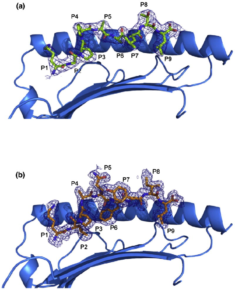
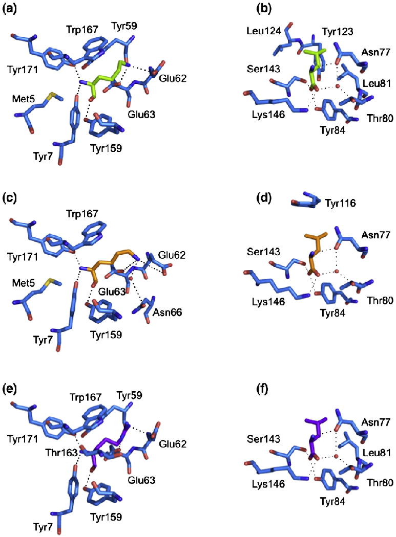
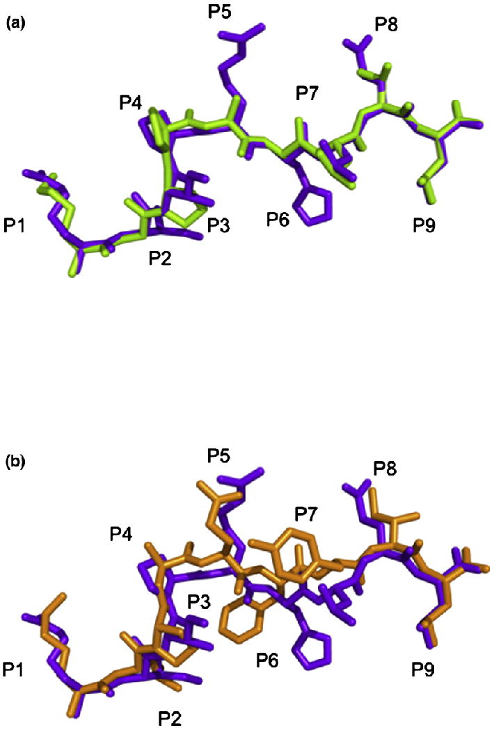
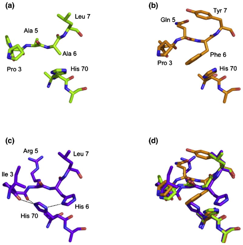
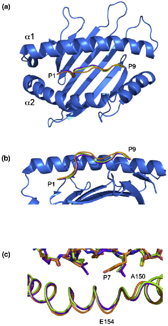
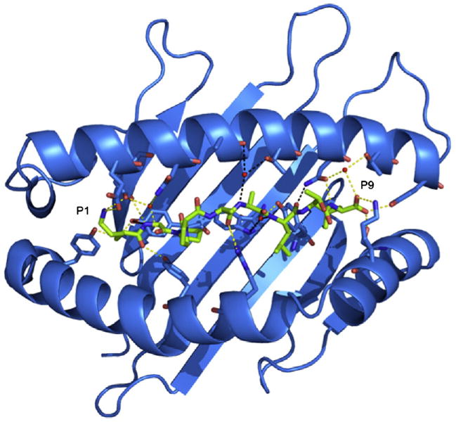
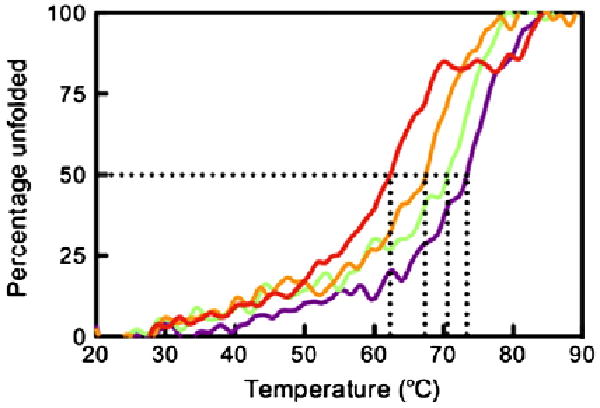
The highly polymorphic major histocompatibility complex class Ia (MHC-Ia) molecules present a broad array of peptides to the clonotypically diverse alphabeta T-cell receptors. In contrast, MHC-Ib molecules exhibit limited polymorphism and bind a more restricted peptide repertoire, in keeping with their major role in innate immunity. Nevertheless, some MHC-Ib molecules do play a role in adaptive immunity. While human leukocyte antigen E (HLA-E), the MHC-Ib molecule, binds a very restricted repertoire of peptides, the peptide binding preferences of HLA-G, the class Ib molecule, are less stringent, although the basis by which HLA-G can bind various peptides is unclear. To investigate how HLA-G can accommodate different peptides, we compared the structure of HLA-G bound to three naturally abundant self-peptides (RIIPRHLQL, KGPPAALTL and KLPQAFYIL) and their thermal stabilities. The conformation of HLA-G(KGPPAALTL) was very similar to that of the HLA-G(RIIPRHLQL) structure. However, the structure of HLA-G(KLPQAFYIL) not only differed in the conformation of the bound peptide but also caused a small shift in the alpha2 helix of HLA-G. Furthermore, the relative stability of HLA-G was observed to be dependent on the nature of the bound peptide. These peptide-dependent effects on the substructure of the monomorphic HLA-G are likely to impact on its recognition by receptors of both innate and adaptive immune systems.


Figures

Fig. 1
Fig. 1
Structure of pHLA-G complexes. (a and b) Side views of the HLA-GKGPPAALTL complex (a) and the HLA-GKLPAQFYIL complex (b) showing 2.4- and 1.7-Å omit maps, respectively (contoured at 1σ). The α2 helix has been removed for clarity.
Fig. 2
Fig. 2
Conserved interactions between peptide and HLA-G. The polar and non-polar contacts of the P1 residue (a, c, e) and the P9 residue (b, d, f) of each pHLA-G complex are shown. (a and b) HLA-GKGPPAALTL. (c and d) HLA-GKLPAQFYIL. (e and f) HLA-GRIIPRHLQL. H-bonds and salt bridges are represented by dashed lines, and water molecules are represented by red spheres.
Fig. 3
Fig. 3
Peptide conformation. (a) Comparison of the conformations of the KGPPAALTL peptide (green) and the RIIPRHLQL peptide (purple) when presented by HLA-G highlighting the similarity between the two peptides. (b) Comparison of the conformations of the KLPAQFYIL peptide (orange) and the RIIPRHLQL peptide (purple) when presented by HLA-G highlighting the central bulge in KLPAQFYIL.
Fig. 4
Fig. 4
Orientation of His70. The orientation of His70 side chain is altered by the presence of a His at P6 of RIIPRHLQL (c) compared with that seen in the presence of KGPPAALTL (a) and KLPAQFYIL (b). An overlay of the three peptides is shown in (d).
Fig. 5
Fig. 5
Comparison of the three pHLA-G complexes. Top view (a) and side view (b) of HLA-G showing the backbones of RIIPRHLQL (purple), KGPPAALTL (green) and KLPAQFYIL (orange). The α2 helix has been removed from (b) for clarity. (c) A 0.9-Å shift in the α2 helix centered at V152 is seen in the HLA-GKLPAQFYIL complex (orange) when compared with HLA-GRIIPRHLQL (purple) and HLA-GKGPPAALTL (green).
Fig. 6
Fig. 6
Conserved peptide contacts. Polar contacts between the KGPPAALTL peptide and HLA-G are represented by dashed lines shown in yellow for conserved contacts and in black for non-conserved contacts. Water molecules are represented by red spheres.
Fig. 7
Fig. 7
Thermal stability of pHLA-G complexes. The ellipticity at 218 nm was measured as temperature increased from 20 to 90 °C. The data were normalised to determine the midpoint of thermal denaturation (Tm). Samples were diluted to 5 and 10 μM in 10 mM Tris, pH 8.0, containing 150 mM NaCl. The complexes were measured twice at both 5 and 10 μM, and the Tm was averaged over the four experiments. The figure is representative of denaturation at 5 μM. Data for HLA-GRLPKDFRIL, HLA-GKLPAQFYIL, HLA-GKGPPAALTL and HLA-GRIIPRHLQL are shown in red, orange, green and purple, respectively.

Similar articles

Cited by

References

    1. Archbold JK, Macdonald WA, Gras S, Ely LK, Miles JJ, Bell MJ, et al. Natural micropolymorphism in human leukocyte antigens provides a basis for genetic control of antigen recognition. J Exp Med. 2009;206:209–219. - PMC - PubMed
    1. Macdonald WA, Purcell AW, Mifsud NA, Ely LK, Williams DS, Chang L, et al. A naturally selected dimorphism within the HLA-B44 supertype alters class I structure, peptide repertoire, and T cell recognition. J Exp Med. 2003;198:679–691. - PMC - PubMed
    1. Tynan FE, Borg NA, Miles JJ, Beddoe T, El-Hassen D, Silins SL, et al. High resolution structures of highly bulged viral epitopes bound to major histocompatibility complex class I: implications for T-cell receptor engagement and T-cell immunodominance. J Biol Chem. 2005;280:23900–23909. - PubMed
    1. Grimsley C, Kawasaki A, Gassner C, Sageshima N, Nose Y, Hatake K, et al. Definitive high resolution typing of HLA-E allelic polymorphisms: identifying potential errors in existing allele data. Tissue Antigens. 2002;60:206–212. - PubMed
    1. Lajoie J, Boivin AA, Jeanneau A, Faucher MC, Roger M. Identification of six new HLA-G alleles with non-coding DNA base changes. Tissue Antigens. 2009;73:379–380. - PubMed

Publication types