. 2010 Apr 2;397(3):647-63.
doi: 10.1016/j.jmb.2010.01.049. Epub 2010 Feb 1.

Crystal structure of the first eubacterial Mre11 nuclease reveals novel features that may discriminate substrates during DNA repair

Affiliations

Crystal structure of the first eubacterial Mre11 nuclease reveals novel features that may discriminate substrates during DNA repair

Debanu Das et al. J Mol Biol. .

Abstract

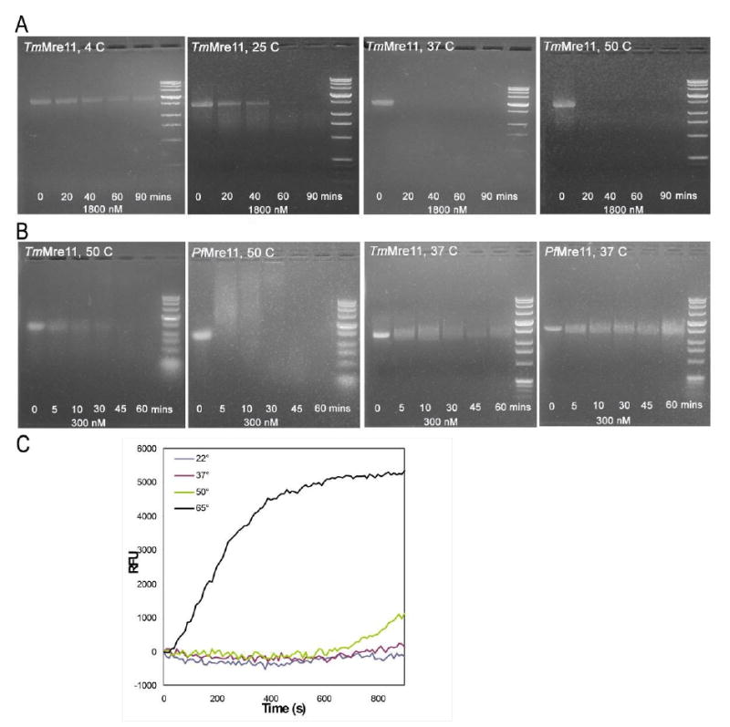
Mre11 nuclease plays a central role in the repair of cytotoxic and mutagenic DNA double-strand breaks. As X-ray structural information has been available only for the Pyrococcus furiosus enzyme (PfMre11), the conserved and variable features of this nuclease across the domains of life have not been experimentally defined. Our crystal structure and biochemical studies demonstrate that TM1635 from Thermotoga maritima, originally annotated as a putative nuclease, is an Mre11 endo/exonuclease (TmMre11) and the first such structure from eubacteria. TmMre11 and PfMre11 display similar overall structures, despite sequence identity in the twilight zone of only approximately 20%. However, they differ substantially in their DNA-specificity domains and in their dimeric organization. Residues in the nuclease domain are highly conserved, but those in the DNA-specificity domain are not. The structural differences likely affect how Mre11 from different organisms recognize and interact with single-stranded DNA, double-stranded DNA and DNA hairpin structures during DNA repair. The TmMre11 nuclease active site has no bound metal ions, but is conserved in sequence and structure with the exception of a histidine that is important in PfMre11 nuclease activity. Nevertheless, biochemical characterization confirms that TmMre11 possesses both endonuclease and exonuclease activities on single-stranded and double-stranded DNA substrates, respectively.


Figures

Fig. 1
Fig. 1. Crystal structure of TmMre11
(A) Stereo ribbon diagram of the TmMre11 monomer color-coded from N-terminus (yellow) to C-terminus (green). TmMre11 is a ladle-shaped molecule with two domains. Helices H1–H8 and β-strands β1-β15 that comprise the Nuclease Domain (nuc_domain) (approximately residues 1-270) and Specificity Domain (spec_domain) (approximately residues 271-324) are indicated. The nuc_domain has a β-sandwich architecture formed by two 6-stranded β-sheets surrounded by α-helices. The spec_domain is formed by a 3-stranded β-sheet and two α-helices. (B) Diagram showing the secondary structure elements of TmMre11 superimposed on its primary sequence, adapted from PDBsum (http://www.ebi.ac.uk/pdbsum), where α-helices are sequentially labeled (H1, H2, H3 etc), β-strands labeled (β1, β2, β3, etc) and β-hairpins by red loops.
Fig. 2
Fig. 2. Comparisons of different Mre11 proteins
(A) Combination of structure- and sequence-based sequence alignment to analyze similarities and differences between Mre11 proteins from T. maritima, P. furiosus, human and S. cerevisiae. The TmMre11 and PfMre11 proteins have been aligned by a structure-based sequence alignment using their 3D coordinates (Protein Data Bank files) as input. This alignment was then used as an anchor for the sequence-based sequence alignment of the HuMre11 and ScMre11. The TmMre11 chain B, which has residues 1-6 disordered, was used since it has all of the catalytic site residues modeled in the structure. Chain A has residues 1-6, but does not have the active site His94. Therefore, in the figure, the alignment for TmMre11 starts after the dots at residue 7. All the proteins show regions of conserved residues in the nuc_domain, which are involved in endo- and exonucleolytic cleavage of DNA. No significant sequence conservation is found in the spec_domain, which suggests that this may lead to differences in recognition of single-stranded DNA, double-stranded DNA and DNA hairpin structures during the DNA repair process in different species. (B) Left panel: The structure of TmMre11 (blue) superimposed on PfMre11 (gray, red and green) reveals their overall similarity. However, the spec_dom of TmMre11 is smaller with 3 β-strands as opposed to 5 β-strands in PfMre11 (gray and red). The nuc_domain of PfMre11 also has an extra insertion (green) that is not found in TmMre11. Right panel: The spec_domain of TmMre11 (blue) and PfMre11 (gray) based on superimposing only by the nuc_domain of the two proteins reveals that the relative orientation of the spec_domain with respect to the nuc_domain is different in the two proteins. Thus, the combination of the smaller spec_domain and slightly different juxtaposition of the two domains in TmMre11 is likely to impact substrate recognition. (C) Comparison of the nuclease active site of TmMre11 (blue) with PfMre11 (gray). The PfMre11 His85 residue (magenta) that is critical in nuclease activity is above the plane of the other residues His206, His208, His10, His173 and Asp49 (all green) that form the rest of the active site and coordinate the metal (orange spheres). In TmMre11, His94 (red) corresponds to the PfMre11 His85 and is positioned differently that could be due to the lack of a metal in the active site. The other active site residues in the TmMre11 are His216, His218, His16, His180 and Asp58 (all cyan). The inset depicts a larger, focused view of the active site.
Fig. 3
Fig. 3. Oligomerization in TmMre11 and PfMre11
(A) The TmMre11 dimer is viewed along the non-crystallographic 2-fold axis of the dimer (blue and green). (B) The PfMre11 dimer (gray and red) is depicted in approximately the same orientation as the TmMre11 dimer, revealing the difference in dimeric assembly as observed in the crystal structures. (C) Details of the dimer interface of TmMre11 are shown. Phe102 (orange) and Phe105 (cyan) in helices H3 and H3′ play a key role in assembly of the dimer interface. His72 (yellow) is also present in the HuMre11. Leu75 (grey), Leu78 (pink), Lys79 (blue) and Ile113 (magenta) are found in TmMre11, PfMre11, HuMre11 and ScMre11. Conservation of these residues at the dimer interface suggests that they may be needed for dimer formation.
Fig. 4
Fig. 4. DNA binding residues in TmMre11
(A) Residues at the nuclease active site (cyan) are conserved between the TmMre11 and PfMre11. Several other TmMre11 residues (Trp22, His61, Ser67, Lys322, Tyr298, Arg26 and Pro27) that are the counterparts of DNA binding residues in PfMre11 are shown. Additionally, other residues in the spec_domain that are conserved in TmMre11 and orthologs from T. petrophila RKU-1, M. piezophila KA3, T. sp. RQ2 and T. neapolitana DSM 4359 and that may be involved in substrate recognition and discrimination are shown. (B) Surface representation of the TmMre11 monomer showing color-coded according to amino acid conservation based on comparison to 374 unique Mre11/SbcD proteins. The highest conservation is seen in the nuc_domain (red spheres) and least conservation in the spec_domain (cyan spheres).
Fig. 5
Fig. 5. Biochemical characterization of TmMre11
TmMre11 was originally annotated in UniProt as a putative nuclease. Assays were performed to verify Mre11 activity. (A) Endonuclease assay using TmMre11 with single-stranded bacteriophage φX174 DNA as substrate. From left to right, the gels are for reactions at 4 °C, 25 °C, 37 °C and 50 °C, respectively. The lanes on each gel, from left to right, are for incubation times of 0 (no enzyme control), 20, 40, 60 and 90 minutes, respectively. The right lane on each gel is the DNA standard. At lower temperatures, the DNA substrate is not substantially cleaved even over a long time period and the corresponding bands remain visible in the gel. At the higher temperatures, the disappearance of the substrate band indicates that endonucleolytic activity has occurred. Thus, TmMre11 is active on ssDNA substrate and the activity is directly proportional to the temperature of the reaction, which is expected for a thermophile. (B) Comparison of the endonuclease activities of TmMre11 and PfMre11 on bacteriophage φX174 at 37 °C and 50 °C. From left to right, the gels are for TmMre11 and PfMre11 at 50 °C followed by the reaction at 37 °C. On each gel, from left to right, the lanes are the control reaction (no enzyme), products at time 5, 10, 30, 45 and 60 minutes and the DNA marker standard. Both enzymes, being thermophilic, display higher activity at 50 °C, with TmMre11 showing slightly higher activity at this temperature. At 37 °C, PfMre11 does not appear to have significant activity in the first 30 minutes of the reaction and TmMre11 has higher activity. (C) Exonuclease assay on double-stranded DNA. The reaction graph depicts, in the y-axis, the RFU (Relative Fluorescence Unit) monitored in the release of 2-aminopurine bases from the 2-aminopurine containing DNA duplex due to exonuclease activity. The x-axis shows the time (in seconds) of the period (15 minutes) over which the reaction was allowed to proceed. TmMre11 shows efficient exonuclease activity at 65 °C and weaker activity at 50 °C. The 50 °C reaction has ∼10% increase in the last 5 minutes and the 65 °C reaction has ∼60% linear increase during that time.
Fig. 6
Fig. 6. Endonuclease assays on TmMre11 mutants
Endonuclease assays were performed at 50 °C with single-stranded bacteriophage φX174 DNA as substrate. The lanes, from left to right, are for incubation times of 0 (no enzyme control), 5, 10, 20, 30 and 40 minutes, respectively. The left lane is the DNA standard. (A) The TmMre11-His94Ser mutant (which most likely interacts with dAMP and corresponds to His85 of PfMre11) results in total loss of function and no cleavage of substrate is observed, demonstrating that this residue is critical for biochemical activity. (B) and (C) Mutation of two of the five putative metal binding residues, His180Ser and His216Ser result in partial loss of function; substrate cleavage appears to occur ∼20 minutes after start of the reaction. (D) The His61Ser mutant (corresponds to His52 in PfMre11, whose primary role is in exonuclease activity, and mutation leads to partial loss of endonuclease activity) shows a partial loss of endonuclease function.

References

    1. Ward JF. DNA damage produced by ionizing radiation in mammalian cells: identities, mechanisms of formation, and reparability. Prog Nucleic Acid Res Mol Biol. 1988;35:95–125. - PubMed
    1. Bierne H, Ehrlich SD, Michel B. Deletions at stalled replication forks occur by two different pathways. EMBO J. 1997;16:3332–3340. - PMC - PubMed
    1. Gellert M. Recent advances in understanding V(D)J recombination. Adv Immunol. 1997;64:39–64. - PubMed
    1. Michel B, Ehrlich SD, Uzest M. DNA double-strand breaks caused by replication arrest. EMBO J. 1997;16:430–438. - PMC - PubMed
    1. Williams RS, Williams JS, Tainer JA. Mre11-Rad50-Nbs1 is a keystone complex connecting DNA repair machinery, double-strand break signaling, and the chromatin template. Biochem Cell Biol. 2007;85:509–520. - PubMed

Publication types

Associated data

LinkOut - more resources