. 2010 May 5;167(2):287-97.
doi: 10.1016/j.neuroscience.2010.01.052. Epub 2010 Feb 1.

Alpha-2 noradrenergic receptor activation inhibits the hyperpolarization-activated cation current (Ih) in neurons of the ventral tegmental area

Affiliations

Alpha-2 noradrenergic receptor activation inhibits the hyperpolarization-activated cation current (Ih) in neurons of the ventral tegmental area

M U Inyushin et al. Neuroscience. .

Abstract

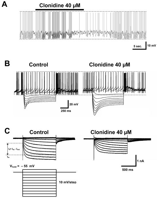
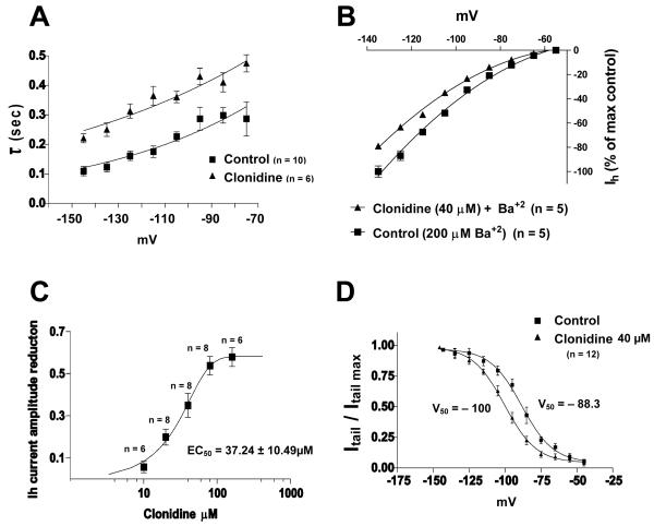
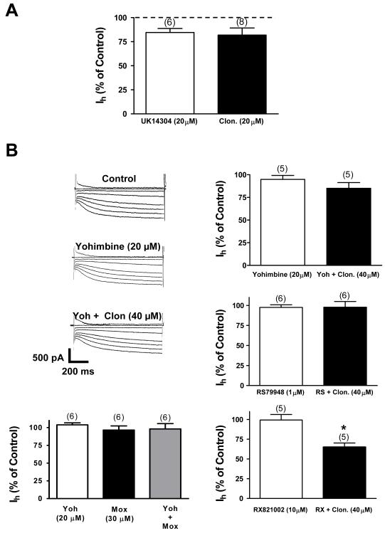
The ventral tegmental area (VTA) is the source of dopaminergic projections innervating cortical structures and ventral forebrain. Dysfunction of this mesocorticolimbic system is critically involved in psychiatric disorders such as addiction and schizophrenia. Changes in VTA dopamine (DA) neuronal activity can alter neurotransmitter release at target regions which modify information processing in the reward circuit. Here we studied the effect of alpha-2 noradrenergic receptor activation on the hyperpolarization-activated cation current (I(h)) in DA neurons of the rat VTA. Brain slice preparations using whole-cell current and voltage-clamp techniques were employed. Clonidine and UK14304 (alpha-2 receptor selective agonists) were found to decrease I(h) amplitude and to slow its rate of activation indicating a negative shift in the current's voltage dependence. Two non-subtype-selective alpha-2 receptor antagonists, yohimbine and RS79948, prevented the effects of alpha-2 receptor activation. RX821002, a noradrenergic antagonist specific for alpha-2A and alpha-2D did not prevent I(h) inhibition. This result suggests that clonidine might be acting via an alpha-2C subtype since this receptor is the most abundant variant in the VTA. Analysis of a second messenger system associated with the alpha-2 receptor revealed that I(h) inhibition is independent of cyclic AMP (cAMP) and resulted from the activation of protein kinase C. It is suggested that the alpha-2 mediated hyperpolarizing shift in I(h) voltage dependence can facilitate the transition from pacemaker firing to afferent-driven burst activity. This transition may play a key role on the changes in synaptic plasticity that occurs in the mesocorticolimbic system under pathological conditions.


Figures

Fig. 1
Fig. 1. Clonidine’s effect on VTA DA cells
A. A 20 seconds bath application of the α-2 noradrenergic agonist clonidine (40 μM) evokes a slight depolarization of the membrane potential in VTA DA cells (1.7 ± 0.4 mV, n = 10). Concomitant with this event there is a significant decrease in the following measurements: action potential amplitude, afterhyperpolarization amplitude and, rate of subthreshold depolarization. There is a significant increase in the interspike interval. B. Current-clamp response of a VTA DA cell to a family of 8 hyperpolarizing dc current pulses of 100 pA each (duration: 1 sec, frequency: 1/9 Hz) in the absence and presence of clonidine. The cell was held at 0 pA. Note that all the effects seen above (part A of the figure) are also seen here. The amplitude of voltage responses under clonidine is increased indicating an augmentation in the cell’s input resistance. Voltage “sag” is also more profound and visibly slowed indicating a shift in the voltage dependence of the underlying Ih. C. In voltage clamp mode, bath application of clonidine reduces the difference between the steady state current (Iss) and the instantaneous current (Iinst) at all command potentials below the holding level (−55 mV), i.e., clonidine inhibits Ih = Iss − Iinst. In the presence of clonidine the membrane current reaches its steady state value at a much slower rate. As indicated by part B of the figure, clonidine’s effects on Ih, membrane potential and firing frequency follow a similar time course.
Fig. 2
Fig. 2. Clonidine causes significant negative shift in Ih voltage dependence and Kir channels are not involved in this effect
A. Monoexponential time constants (τ) vs membrane potential plot indicates that clonidine (40μM) slows down Ih activation upon membrane hyperpolarization, i.e., greater τ. B. Normalized current-voltage curve showing that barium (Ba+2) does not alter clonidine’s effect on Ih, implying that Kir channels are not involved. C. Dose-response curve of clonidine’s reduction of Ih current amplitude at −125 mV, illustrating that there is an increase inhibition at higher concentrations. D. Activation curve showing that clonidine (40μM) produces a negative shift (−11.7 mV) in Ih voltage dependence (n = 12 is the same in both groups).
Fig. 3
Fig. 3. Clonidine inhibits Ih by activation of the α-2 noradrenergic receptor
A. Application of the α-2 noradrenergic agonist UK14304 (20 μM) causes Ih inhibition in VTA DA cells. This inhibition is similar to the one evoked by 20 μM of clonidine (P > 0.05). The numbers above the bars indicate the number of cells that were used for each treatment. B. Current traces show that yohimbine, a selective non-subtype α-2 noradrenergic antagonist, blocks clonidine’s inhibitory effect. To the right, bar graphs summarize the effects of yohimbine. RS79948, another selective non-subtype- α-2 noradrenergic antagonist, also blocks clonidine’s action. Moxonidine (Mox) in the presence of yohimbine activates preferentially imidazoline receptors. The Ih was not decreased under this condition or under moxonidine alone. It seems that clonidine’s action is mediated by activation of α-2 receptors and do not involve an imidazoline receptor component. RX821002 (10 μM), an antagonist for α-2A and α-2D subtypes did not abolish clonidine’s evoked Ih inhibition. This inhibition was statistically different from that of RX 821002 alone (P < 0.05). Consequently, it appears that clonidine’s action is mediated by a specific α-2C receptor subtype. In all graphs the numbers above the bars indicate the number of cells that were used for each treatment.
Fig. 4
Fig. 4. Activation of α-2 noradrenergic receptor stimulates PKC in a calcium insensitive manner
A. The bar graph shows that loading the patch pipette with 3 mM of cAMP does not block clonidine’s effect on Ih current. Minutes within the bar indicate the exposition time to cAMP. Note that there is not a significant difference between clonidine alone and clonidine plus cAMP (P > 0.05). The numbers above the bars indicate the number of cells that were used for each treatment. Asterisk denotes P < 0.05. B. The bar graph shows that bath application of 5 μM of the PKC activator phorbol 12,13 diacetate (PDA) inhibits Ih and occludes clonidine’s effect. Additionally, bath application of 1 μM of the PKC antagonist chelerythrine blocks clonidine’s action indicating that PKC is necessary for clonidine-induced Ih inhibition in VTA DA cells. Note that there is not a significant difference between clonidine alone and clonidine plus PDA (P > 0.05). The numbers above the bars indicate the number of cells that were used for each treatment. C. The bar graph shows that loading the patch pipette with 10 mM of the Ca2+ chelator BAPTA increases Ih amplitude. Minutes within the bar indicate the exposition time to BAPTA. This treatment does not block clonidine’s effect on Ih. Note that there is not a significant difference between clonidine alone and clonidine plus BAPTA (P > 0.05). The numbers above the bars indicate the number of cells that were used for each treatment. Asterisk denotes P < 0.05.

Similar articles

Cited by

References

    1. Aghajanian G, Bunney BC. In: Usdin E, Snyder SH, editors. Central dopaminergic neurons: Neurophysiologic identification and responses to drugs; Frontiers in catecholamine research; proceedings; Elmsford, N.Y., Pergamon Press. 1973.pp. 643–648.
    1. Alagem N, Dvir M, Reuveny E. Mechanism of Ba(2+) block of a mouse inwardly rectifying K+ channel: differential contribution by two discrete residues. J Physiol. 2001;534:381–393. - PMC - PubMed
    1. Arencibia-Albite F, Paladini C, Williams JT, Jiménez-Rivera CA. Noradrenergic modulation of the hyperpolarization-activated cation current (Ih) in dopamine neurons of the ventral tegmental area. Neuroscience. 2007;149:303–314. - PMC - PubMed
    1. Aston-Jones G, Kalivas P. Brain norepinephrine rediscovered in addiction research. Biol Psychiatry. 2008;63:1005–1006. - PMC - PubMed
    1. Berger T, Senn W, Luscher RL. Hyperpolarization-activated current Ih disconnects somatic and dendritic spike initiation zones in layer V pyramidal neurons. J. Neurophysiol. 2003;90:2428–2437. - PubMed

Publication types